|
| ||||||
| Protein Homology/analogY Recognition Engine V 2.2 | |||||||
|
|
|
| ||||||
| Protein Homology/analogY Recognition Engine V 2.2 | |||||||
|
| Fold library id | PDB Header | Molecule | Title |
|---|---|---|---|
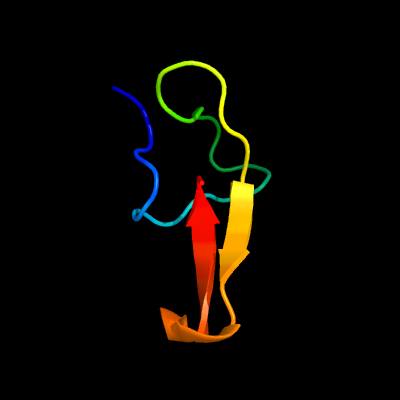
| c2mquA_ | PDB header: toxin | Chain: A: PDB Molecule: neurotoxin hm-3; | PDBTitle: spatial structure of hm-3, a membrane-active spider toxin affecting2 sodium channels |
| Added to library: Sat Nov 8 08:16:51 2014 | |
| Links to external resources | |
|---|---|
| 1 | . | . | . | . | . | . | . | . | 10 | . | . | . | . | . | . | . | . | . | 20 | . | . | . | . | . | . | . | . | . | 30 | . | . | . | . | . | |
| Sequence | G | C | I | A | K | N | K | E | C | A | W | F | S | G | E | W | C | C | G | A | L | S | C | K | Y | S | I | K | R | N | L | K | I | C | V |
| Predicted secondary structure |  | | | |  | | | | | | | | | | | | | | | | |||||||||||||||
| SS confidence | |||||||||||||||||||||||||||||||||||
| Known secondary structure (DSSP) | B | T | T | S | B | T | T | S | S | B | S | T | T | | | | | G | G | G | T | | | | |
| Download: | PDB structure | FASTA sequence |
Phyre is now FREE for commercial users! All images and data generated by Phyre2 are free to use in any publication with acknowledgement Accessibility Statement
| ||||||||||||||||||||||||