|
| ||||||
| Protein Homology/analogY Recognition Engine V 2.2 | |||||||
|
|
|
| ||||||
| Protein Homology/analogY Recognition Engine V 2.2 | |||||||
|
| Fold library id | PDB Header | Molecule | Title |
|---|---|---|---|
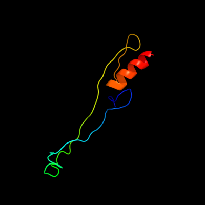
| c2hgj3_ | PDB header: ribosome | Chain: 3: PDB Molecule: 50s ribosomal protein l31; | PDBTitle: crystal structure of the 70s thermus thermophilus ribosome showing how2 the 16s 3'-end mimicks mrna e and p codons. this entry 2hgj contains3 50s ribosomal subunit. the 30s ribosomal subunit can be found in pdb4 entry 2hgi. |
| Added to library: Thu Jan 10 05:03:28 2013 | |
| Links to external resources | |
|---|---|
| 1 | . | . | . | . | . | . | . | . | 10 | . | . | . | . | . | . | . | . | . | 20 | . | . | . | . | . | . | . | . | . | 30 | . | . | . | . | . | . | . | . | . | 40 | . | . | . | . | . | . | . | . | . | 50 | . | . | . | . | . | . | . | . | . | 60 | . | . | . | . | . | . | . | . | . | 70 | |
| Sequence | M | K | E | G | I | H | P | K | L | V | P | A | R | I | I | C | G | C | G | N | V | I | E | T | Y | S | T | K | P | E | I | Y | V | E | V | C | S | K | C | H | P | F | Y | T | G | Q | Q | R | F | V | D | T | E | G | R | V | E | R | F | Q | R | R | Y | G | D | S | Y | R | K | G |
| Predicted secondary structure |  | | | | | | |  | | | | | | | | | | | | | | | | | | | |  | | | | | | | | | | | | | | | | | ||||||||||||||||||||||||||
| SS confidence | ||||||||||||||||||||||||||||||||||||||||||||||||||||||||||||||||||||||
| Known secondary structure (DSSP) | S | S | S | S | | | B | G | G | G | T | B | | | | | | | S | S | S | S | | | | | | | | | | | | | | | | | | . | ||||||||||||||||||||||||||||||
| Sequence | R | |||||||||||||||||||||||||||||||||||||||||||||||||||||||||||||||||||||
| Predicted secondary structure | ||||||||||||||||||||||||||||||||||||||||||||||||||||||||||||||||||||||
| SS confidence | ||||||||||||||||||||||||||||||||||||||||||||||||||||||||||||||||||||||
| Known secondary structure (DSSP) |
| Download: | PDB structure | FASTA sequence |
Phyre is now FREE for commercial users! All images and data generated by Phyre2 are free to use in any publication with acknowledgement Accessibility Statement
| ||||||||||||||||||||||||