Phyre Home Retrieve Phyre Job Id
Emaill.a.kelley@imperial.ac.uk
DescriptionP77427
DateThu Jan 5 12:29:11 GMT 2012
Unique Job IDff499a8ef9317652

Summary 

Top model
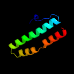
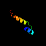
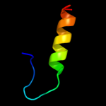
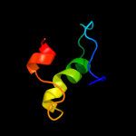
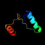
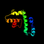
Image coloured by rainbow N → C terminus
Model (left) based on template d2cwla1
Top template information
Fold:Ferritin-like
Superfamily:Ferritin-like
Family:Manganese catalase (T-catalase)
Confidence and coverage
Confidence: 34.0% Coverage: 28%
66 residues ( 28% of your sequence) have been modelled with 34.0% confidence by the single highest scoring template.
You may wish to submit your sequence to Phyrealarm. This will automatically scan your sequence every week for new potential templates as they appear in the Phyre2 library.
Please note: You must be registered and logged in to use Phyrealarm.
3D viewing
Interactive 3D view in Jmol

Sequence analysis 

Secondary structure and disorder prediction 

   1........10.........20.........30.........40.........50.........60
Sequence  MNKEEQYLLFALSAPMEILNQGCKPAHDSPKMYTGIKEFELSSSWGINNRDDLIQTIYQM
Secondary structure 


SS confidence 


Disorder  ??


?

?


Disorder confidence 


   .........70.........80.........90.........100.........110.........120
Sequence  TDDGHANDLAGLYLTWHRSSPEEWKALIAGGSERGLIYTQFVAQTAMCCGEGGIKAWDYV
Secondary structure 


SS confidence 


Disorder 


Disorder confidence 


   .........130.........140.........150.........160.........170.........180
Sequence  RMGFLSRVGVLNKWLTEEESLWLQSRVYVRAHHYYHSWMHYFSAYSLGRLYWQSSQCEDN
Secondary structure 


SS confidence 


Disorder 


?????
Disorder confidence 


   .........190.........200.........210.........220.........230.....
Sequence  TSLREALTLYKYDSAGSRMFEELAAGSDRFYATLPWQPLTVQSECPVTLKDVSDL
Secondary structure 


SS confidence 


Disorder  ?


?????


?

????
Disorder confidence 


Confidence Key
High(9)                    Low (0)
?Disordered
Alpha helix
Beta strand

Domain analysis 

Hover over an aligned region to see model and summary info

Please note, only up to the top 20 hits are modelled to reduce computer load

RankAligned region

PDB 2cwl chain A domain 1

3D model

Region: 12 - 77
Aligned: 66
Modelled: 66
Confidence: 34.0%
Identity: 8%
Fold: Ferritin-like
Superfamily: Ferritin-like
Family: Manganese catalase (T-catalase)

Phyre2

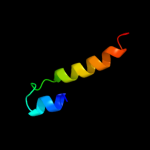
PDB 2job chain A

3D model

Region: 124 - 145
Aligned: 22
Modelled: 22
Confidence: 25.6%
Identity: 14%
PDB header:lipid binding protein
Chain: A: PDB Molecule:antilipopolysaccharide factor;
PDBTitle: solution structure of an antilipopolysaccharide factor from2 shrimp and its possible lipid a binding site

Phyre2

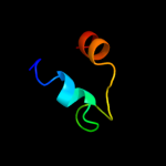
PDB 1jog chain A

3D model

Region: 124 - 156
Aligned: 33
Modelled: 33
Confidence: 24.7%
Identity: 9%
Fold: Four-helical up-and-down bundle
Superfamily: Nucleotidyltransferase substrate binding subunit/domain
Family: Family 1 bi-partite nucleotidyltransferase subunit

Phyre2

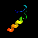
PDB 1jkv chain A

3D model

Region: 12 - 78
Aligned: 67
Modelled: 67
Confidence: 23.8%
Identity: 4%
Fold: Ferritin-like
Superfamily: Ferritin-like
Family: Manganese catalase (T-catalase)

Phyre2

PDB 1wwp chain A

3D model

Region: 124 - 156
Aligned: 33
Modelled: 33
Confidence: 20.3%
Identity: 18%
PDB header:structural genomics, unknown function
Chain: A: PDB Molecule:hypothetical protein ttha0636;
PDBTitle: crystal structure of ttk003001694 from thermus thermophilus2 hb8

Phyre2

PDB 1wty chain A

3D model

Region: 124 - 156
Aligned: 33
Modelled: 33
Confidence: 20.1%
Identity: 15%
Fold: Four-helical up-and-down bundle
Superfamily: Nucleotidyltransferase substrate binding subunit/domain
Family: Family 1 bi-partite nucleotidyltransferase subunit

Phyre2

PDB 2lhu chain A

3D model

Region: 29 - 57
Aligned: 24
Modelled: 29
Confidence: 19.0%
Identity: 17%
PDB header:structural protein
Chain: A: PDB Molecule:mybpc3 protein;
PDBTitle: structural insight into the unique cardiac myosin binding protein-c2 motif: a partially folded domain

Phyre2

PDB 1tz7 chain A domain 1

3D model

Region: 40 - 66
Aligned: 27
Modelled: 27
Confidence: 17.0%
Identity: 19%
Fold: TIM beta/alpha-barrel
Superfamily: (Trans)glycosidases
Family: Amylase, catalytic domain

Phyre2

PDB 1esw chain A

3D model

Region: 40 - 66
Aligned: 27
Modelled: 27
Confidence: 14.9%
Identity: 11%
Fold: TIM beta/alpha-barrel
Superfamily: (Trans)glycosidases
Family: Amylase, catalytic domain

Phyre2

PDB 2adv chain B

3D model

Region: 203 - 213
Aligned: 11
Modelled: 11
Confidence: 14.6%
Identity: 18%
PDB header:hydrolase
Chain: B: PDB Molecule:glutaryl 7- aminocephalosporanic acid acylase;
PDBTitle: crystal structures of glutaryl 7-aminocephalosporanic acid acylase:2 mutational study of activation mechanism

Phyre2

PDB 2oa4 chain A domain 1

3D model

Region: 106 - 142
Aligned: 36
Modelled: 37
Confidence: 12.5%
Identity: 19%
Fold: DNA/RNA-binding 3-helical bundle
Superfamily: TrpR-like
Family: SPO1678-like

Phyre2

PDB 2gyk chain B domain 1

3D model

Region: 146 - 167
Aligned: 22
Modelled: 22
Confidence: 12.2%
Identity: 14%
Fold: His-Me finger endonucleases
Superfamily: His-Me finger endonucleases
Family: HNH-motif

Phyre2

PDB 2jrt chain A

3D model

Region: 106 - 142
Aligned: 36
Modelled: 37
Confidence: 11.1%
Identity: 19%
PDB header:structural genomics, unknown function
Chain: A: PDB Molecule:uncharacterized protein;
PDBTitle: nmr solution structure of the protein coded by gene2 rhos4_12090 of rhodobacter sphaeroides. northeast3 structural genomics target rhr5

Phyre2

PDB 1e8o chain A

3D model

Region: 155 - 164
Aligned: 10
Modelled: 10
Confidence: 9.9%
Identity: 30%
Fold: Signal recognition particle alu RNA binding heterodimer, SRP9/14
Superfamily: Signal recognition particle alu RNA binding heterodimer, SRP9/14
Family: Signal recognition particle alu RNA binding heterodimer, SRP9/14

Phyre2

PDB 2fxh chain B

3D model

Region: 178 - 235
Aligned: 58
Modelled: 58
Confidence: 7.0%
Identity: 17%
PDB header:oxidoreductase
Chain: B: PDB Molecule:catalase-peroxidase protein;
PDBTitle: crystal structure of katg at ph 6.5

Phyre2

PDB 2b2q chain B

3D model

Region: 178 - 235
Aligned: 58
Modelled: 58
Confidence: 7.0%
Identity: 17%
PDB header:oxidoreductase
Chain: B: PDB Molecule:catalase-peroxidase;
PDBTitle: crystal structure of native catalase-peroxidase katg at2 ph7.5

Phyre2

PDB 3rdw chain B

3D model

Region: 45 - 91
Aligned: 45
Modelled: 47
Confidence: 6.9%
Identity: 11%
PDB header:oxidoreductase
Chain: B: PDB Molecule:putative arsenate reductase;
PDBTitle: putative arsenate reductase from yersinia pestis

Phyre2

PDB 1x1n chain A domain 1

3D model

Region: 40 - 66
Aligned: 27
Modelled: 27
Confidence: 6.8%
Identity: 11%
Fold: TIM beta/alpha-barrel
Superfamily: (Trans)glycosidases
Family: Amylase, catalytic domain

Phyre2

PDB 2p12 chain A domain 1

3D model

Region: 128 - 157
Aligned: 30
Modelled: 30
Confidence: 6.8%
Identity: 3%
Fold: FomD barrel-like
Superfamily: FomD-like
Family: FomD-like

Phyre2

PDB 2jg6 chain A

3D model

Region: 103 - 205
Aligned: 103
Modelled: 103
Confidence: 6.2%
Identity: 14%
PDB header:hydrolase
Chain: A: PDB Molecule:dna-3-methyladenine glycosidase;
PDBTitle: crystal structure of a 3-methyladenine dna glycosylase i2 from staphylococcus aureus

Phyre2
1

d2cwla1
2

c2jobA_
3

d1joga_
4

d1jkva_
5

c1wwpA_
6

d1wtya_
7

c2lhuA_
8

d1tz7a1
9

d1eswa_
10

c2advB_
11

d2oa4a1
12

d2gykb1
13

c2jrtA_
14

d1e8oa_
15

c2fxhB_
16

c2b2qB_
17

c3rdwB_
18

d1x1na1
19

d2p12a1
20

c2jg6A_
21


22


23


24


25


26


27


28


29


Detailed template information 

#
Template Alignment Coverage3D Model Confidence
% i.d. Template Information
1d2cwla1


34.0 8 Fold:Ferritin-like
Superfamily:Ferritin-like
Family:Manganese catalase (T-catalase)
2c2jobA_


25.6 14 PDB header:lipid binding protein
Chain: A: PDB Molecule:antilipopolysaccharide factor;
PDBTitle: solution structure of an antilipopolysaccharide factor from2 shrimp and its possible lipid a binding site
3d1joga_


24.7 9 Fold:Four-helical up-and-down bundle
Superfamily:Nucleotidyltransferase substrate binding subunit/domain
Family:Family 1 bi-partite nucleotidyltransferase subunit
4d1jkva_


23.8 4 Fold:Ferritin-like
Superfamily:Ferritin-like
Family:Manganese catalase (T-catalase)
5c1wwpA_


20.3 18 PDB header:structural genomics, unknown function
Chain: A: PDB Molecule:hypothetical protein ttha0636;
PDBTitle: crystal structure of ttk003001694 from thermus thermophilus2 hb8
6d1wtya_


20.1 15 Fold:Four-helical up-and-down bundle
Superfamily:Nucleotidyltransferase substrate binding subunit/domain
Family:Family 1 bi-partite nucleotidyltransferase subunit
7c2lhuA_


19.0 17 PDB header:structural protein
Chain: A: PDB Molecule:mybpc3 protein;
PDBTitle: structural insight into the unique cardiac myosin binding protein-c2 motif: a partially folded domain
8d1tz7a1


17.0 19 Fold:TIM beta/alpha-barrel
Superfamily:(Trans)glycosidases
Family:Amylase, catalytic domain
9d1eswa_


14.9 11 Fold:TIM beta/alpha-barrel
Superfamily:(Trans)glycosidases
Family:Amylase, catalytic domain
10c2advB_


14.6 18 PDB header:hydrolase
Chain: B: PDB Molecule:glutaryl 7- aminocephalosporanic acid acylase;
PDBTitle: crystal structures of glutaryl 7-aminocephalosporanic acid acylase:2 mutational study of activation mechanism
11d2oa4a1


12.5 19 Fold:DNA/RNA-binding 3-helical bundle
Superfamily:TrpR-like
Family:SPO1678-like
12d2gykb1


12.2 14 Fold:His-Me finger endonucleases
Superfamily:His-Me finger endonucleases
Family:HNH-motif
13c2jrtA_


11.1 19 PDB header:structural genomics, unknown function
Chain: A: PDB Molecule:uncharacterized protein;
PDBTitle: nmr solution structure of the protein coded by gene2 rhos4_12090 of rhodobacter sphaeroides. northeast3 structural genomics target rhr5
14d1e8oa_


9.9 30 Fold:Signal recognition particle alu RNA binding heterodimer, SRP9/14
Superfamily:Signal recognition particle alu RNA binding heterodimer, SRP9/14
Family:Signal recognition particle alu RNA binding heterodimer, SRP9/14
15c2fxhB_


7.0 17 PDB header:oxidoreductase
Chain: B: PDB Molecule:catalase-peroxidase protein;
PDBTitle: crystal structure of katg at ph 6.5
16c2b2qB_


7.0 17 PDB header:oxidoreductase
Chain: B: PDB Molecule:catalase-peroxidase;
PDBTitle: crystal structure of native catalase-peroxidase katg at2 ph7.5
17c3rdwB_


6.9 11 PDB header:oxidoreductase
Chain: B: PDB Molecule:putative arsenate reductase;
PDBTitle: putative arsenate reductase from yersinia pestis
18d1x1na1


6.8 11 Fold:TIM beta/alpha-barrel
Superfamily:(Trans)glycosidases
Family:Amylase, catalytic domain
19d2p12a1


6.8 3 Fold:FomD barrel-like
Superfamily:FomD-like
Family:FomD-like
20c2jg6A_


6.2 14 PDB header:hydrolase
Chain: A: PDB Molecule:dna-3-methyladenine glycosidase;
PDBTitle: crystal structure of a 3-methyladenine dna glycosylase i2 from staphylococcus aureus
21c1dpuA_


not modelled 6.0 18 PDB header:dna binding protein
Chain: A: PDB Molecule:replication protein a (rpa32) c-terminal domain;
PDBTitle: solution structure of the c-terminal domain of human rpa322 complexed with ung2(73-88)
22d1dpua_


not modelled 6.0 18 Fold:DNA/RNA-binding 3-helical bundle
Superfamily:"Winged helix" DNA-binding domain
Family:C-terminal domain of RPA32
23c2k5eA_


not modelled 6.0 21 PDB header:structural genomics, unknown function
Chain: A: PDB Molecule:uncharacterized protein;
PDBTitle: solution structure of putative uncharacterized protein2 gsu1278 from methanocaldococcus jannaschii, northeast3 structural genomics consortium (nesg) target gsr195
24c3dmlA_


not modelled 5.9 15 PDB header:oxidoreductase
Chain: A: PDB Molecule:putative uncharacterized protein;
PDBTitle: crystal structure of the periplasmic thioredoxin soxs from2 paracoccus pantotrophus (reduced form)
25d1jvaa2


not modelled 5.7 32 Fold:Homing endonuclease-like
Superfamily:Homing endonucleases
Family:Intein endonuclease
26c3gkxB_


not modelled 5.5 12 PDB header:structural genomics, unknown function
Chain: B: PDB Molecule:putative arsc family related protein;
PDBTitle: crystal structure of putative arsc family related protein from2 bacteroides fragilis
27d2gsca1


not modelled 5.5 6 Fold:Bromodomain-like
Superfamily:IVS-encoded protein-like
Family:IVS-encoded protein-like
28d1nkua_


not modelled 5.4 6 Fold:DNA-glycosylase
Superfamily:DNA-glycosylase
Family:3-Methyladenine DNA glycosylase I (Tag)
29d1fsha_


not modelled 5.2 21 Fold:DNA/RNA-binding 3-helical bundle
Superfamily:"Winged helix" DNA-binding domain
Family:DEP domain

Binding site prediction 

Due to computational demand, binding site predictions are not run for batch jobs

If you want to predict binding sites, please manually submit your model of choice to 3DLigandSite


Phyre is for academic use only

Please cite: Protein structure prediction on the web: a case study using the Phyre server
Kelley LA and Sternberg MJE. Nature Protocols 4, 363 - 371 (2009) [pdf] [Import into BibTeX]
 
If you use the binding site predictions from 3DLigandSite, please also cite:
3DLigandSite: predicting ligand-binding sites using similar structures.
Wass MN, Kelley LA and Sternberg MJ Nucleic Acids Research 38, W469-73 (2010) [PubMed]
 
© Structural Bioinformatics Group
Imperial College London
Lawrence Kelley, Benjamin Jefferys
Disclaimer
Terms and Conditions
Component software
Template detection: HHpred 1.51
Secondary structure prediction: Psi-pred 2.5
Disorder prediction: Disopred 2.4
Transmembrane prediction: Memsat_SVM
Multi-template modelling and ab initio: Poing 1.0