| 1 |
|
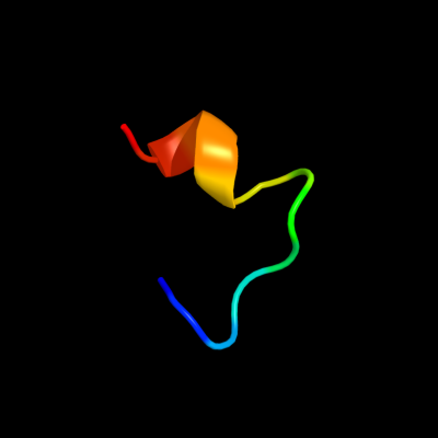
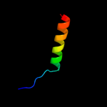
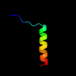
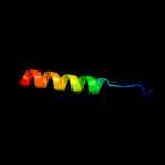
PDB 2ke4 chain A
Region: 37 - 49
Aligned: 13
Modelled: 13
Confidence: 43.0%
Identity: 23%
PDB header:membrane protein
Chain: A: PDB Molecule:cdc42-interacting protein 4;
PDBTitle: the nmr structure of the tc10 and cdc42 interacting domain2 of cip4
Phyre2
| 2 |
|
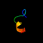
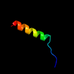
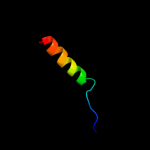
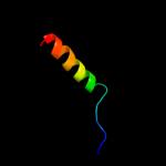
PDB 3bz2 chain L
Region: 39 - 63
Aligned: 25
Modelled: 25
Confidence: 6.6%
Identity: 24%
PDB header:electron transport
Chain: L: PDB Molecule:photosystem ii reaction center protein l;
PDBTitle: crystal structure of cyanobacterial photosystem ii (part 22 of 2). this file contains second monomer of psii dimer
Phyre2
| 3 |
|
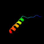
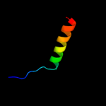
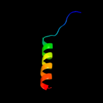
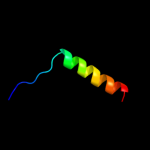
PDB 2axt chain L
Region: 39 - 63
Aligned: 25
Modelled: 25
Confidence: 6.6%
Identity: 24%
PDB header:electron transport
Chain: L: PDB Molecule:photosystem ii reaction center l protein;
PDBTitle: crystal structure of photosystem ii from thermosynechococcus elongatus
Phyre2
| 4 |
|
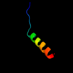
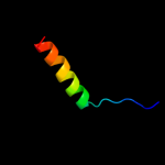
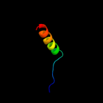
PDB 3prr chain L
Region: 39 - 63
Aligned: 25
Modelled: 25
Confidence: 6.6%
Identity: 24%
PDB header:photosynthesis
Chain: L: PDB Molecule:photosystem ii reaction center protein l;
PDBTitle: crystal structure of cyanobacterial photosystem ii in complex with2 terbutryn (part 2 of 2). this file contains second monomer of psii3 dimer
Phyre2
| 5 |
|
PDB 1s5l chain L
Region: 39 - 63
Aligned: 25
Modelled: 25
Confidence: 6.6%
Identity: 24%
PDB header:photosynthesis
Chain: L: PDB Molecule:photosystem ii reaction center l protein;
PDBTitle: architecture of the photosynthetic oxygen evolving center
Phyre2
| 6 |
|
PDB 3kzi chain L
Region: 39 - 63
Aligned: 25
Modelled: 25
Confidence: 6.6%
Identity: 24%
PDB header:electron transport
Chain: L: PDB Molecule:photosystem ii reaction center protein l;
PDBTitle: crystal structure of monomeric form of cyanobacterial photosystem ii
Phyre2
| 7 |
|
PDB 2axt chain L
Region: 39 - 63
Aligned: 25
Modelled: 25
Confidence: 6.6%
Identity: 24%
PDB header:electron transport
Chain: L: PDB Molecule:photosystem ii reaction center l protein;
PDBTitle: crystal structure of photosystem ii from thermosynechococcus elongatus
Phyre2
| 8 |
|
PDB 3prq chain L
Region: 39 - 63
Aligned: 25
Modelled: 25
Confidence: 6.6%
Identity: 24%
PDB header:photosynthesis
Chain: L: PDB Molecule:photosystem ii reaction center protein l;
PDBTitle: crystal structure of cyanobacterial photosystem ii in complex with2 terbutryn (part 1 of 2). this file contains first monomer of psii3 dimer
Phyre2
| 9 |
|
PDB 3bz1 chain L
Region: 39 - 63
Aligned: 25
Modelled: 25
Confidence: 6.6%
Identity: 24%
PDB header:electron transport
Chain: L: PDB Molecule:photosystem ii reaction center protein l;
PDBTitle: crystal structure of cyanobacterial photosystem ii (part 12 of 2). this file contains first monomer of psii dimer
Phyre2
| 10 |
|
PDB 3a0h chain L
Region: 39 - 63
Aligned: 25
Modelled: 25
Confidence: 6.6%
Identity: 24%
PDB header:electron transport
Chain: L: PDB Molecule:photosystem ii reaction center protein l;
PDBTitle: crystal structure of i-substituted photosystem ii complex
Phyre2
| 11 |
|
PDB 1s5l chain L
Region: 39 - 63
Aligned: 25
Modelled: 25
Confidence: 6.6%
Identity: 24%
PDB header:photosynthesis
Chain: L: PDB Molecule:photosystem ii reaction center l protein;
PDBTitle: architecture of the photosynthetic oxygen evolving center
Phyre2
| 12 |
|
PDB 3a0h chain L
Region: 39 - 63
Aligned: 25
Modelled: 25
Confidence: 6.6%
Identity: 24%
PDB header:electron transport
Chain: L: PDB Molecule:photosystem ii reaction center protein l;
PDBTitle: crystal structure of i-substituted photosystem ii complex
Phyre2
| 13 |
|
PDB 3arc chain L
Region: 39 - 63
Aligned: 25
Modelled: 25
Confidence: 6.6%
Identity: 24%
PDB header:electron transport, photosynthesis
Chain: L: PDB Molecule:photosystem ii reaction center protein l;
PDBTitle: crystal structure of oxygen-evolving photosystem ii at 1.9 angstrom2 resolution
Phyre2
| 14 |
|
PDB 3a0b chain L
Region: 39 - 63
Aligned: 25
Modelled: 25
Confidence: 6.6%
Identity: 24%
PDB header:electron transport
Chain: L: PDB Molecule:photosystem ii reaction center protein l;
PDBTitle: crystal structure of br-substituted photosystem ii complex
Phyre2
| 15 |
|
PDB 2axt chain L domain 1
Region: 39 - 63
Aligned: 25
Modelled: 25
Confidence: 6.6%
Identity: 24%
Fold: Single transmembrane helix
Superfamily: Photosystem II reaction center protein L, PsbL
Family: PsbL-like
Phyre2
| 16 |
|
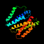
PDB 2gfp chain A
Region: 9 - 227
Aligned: 219
Modelled: 219
Confidence: 6.6%
Identity: 9%
PDB header:membrane protein
Chain: A: PDB Molecule:multidrug resistance protein d;
PDBTitle: structure of the multidrug transporter emrd from2 escherichia coli
Phyre2
| 17 |
|
PDB 3a0b chain L
Region: 39 - 63
Aligned: 25
Modelled: 25
Confidence: 6.4%
Identity: 24%
PDB header:electron transport
Chain: L: PDB Molecule:photosystem ii reaction center protein l;
PDBTitle: crystal structure of br-substituted photosystem ii complex
Phyre2
| 18 |
|
PDB 3arc chain L
Region: 41 - 63
Aligned: 23
Modelled: 23
Confidence: 5.7%
Identity: 26%
PDB header:electron transport, photosynthesis
Chain: L: PDB Molecule:photosystem ii reaction center protein l;
PDBTitle: crystal structure of oxygen-evolving photosystem ii at 1.9 angstrom2 resolution
Phyre2