Phyre Home Retrieve Phyre Job Id
Emaill.a.kelley@imperial.ac.uk
DescriptionP0ADC8
DateThu Jan 5 11:20:37 GMT 2012
Unique Job IDfd59ffe752cf57cd

Summary 

Top model
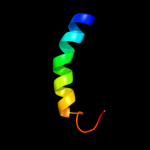
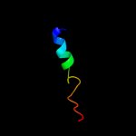
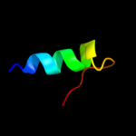
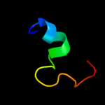
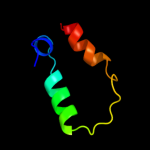
Image coloured by rainbow N → C terminus
Model (left) based on template d1i1rb_
Top template information
Fold:4-helical cytokines
Superfamily:4-helical cytokines
Family:Long-chain cytokines
Confidence and coverage
Confidence: 38.1% Coverage: 37%
25 residues ( 37% of your sequence) have been modelled with 38.1% confidence by the single highest scoring template.
You may wish to submit your sequence to Phyrealarm. This will automatically scan your sequence every week for new potential templates as they appear in the Phyre2 library.
Please note: You must be registered and logged in to use Phyrealarm.
3D viewing
Interactive 3D view in Jmol

Sequence analysis 

Secondary structure and disorder prediction 

   1........10.........20.........30.........40.........50.........60
Sequence  MFGNLGQAKKYLGQAAKMLIGIPDYDNYVEHMKTNHPDKPYMSYEEFFRERQNARYGGDG
Secondary structure 


SS confidence 


Disorder  ??


??????
Disorder confidence 


   .......
Sequence  KGGMRCC
Secondary structure 


SS confidence 


Disorder  ???????
Disorder confidence 


Confidence Key
High(9)                    Low (0)
?Disordered
Alpha helix
Beta strand

Domain analysis 

Hover over an aligned region to see model and summary info

Please note, only up to the top 20 hits are modelled to reduce computer load

RankAligned region

PDB 1i1r chain B

3D model

Region: 17 - 41
Aligned: 25
Modelled: 25
Confidence: 38.1%
Identity: 12%
Fold: 4-helical cytokines
Superfamily: 4-helical cytokines
Family: Long-chain cytokines

Phyre2

PDB 2knc chain B

3D model

Region: 44 - 63
Aligned: 20
Modelled: 20
Confidence: 28.4%
Identity: 20%
PDB header:cell adhesion
Chain: B: PDB Molecule:integrin beta-3;
PDBTitle: platelet integrin alfaiib-beta3 transmembrane-cytoplasmic2 heterocomplex

Phyre2

PDB 3g9w chain C

3D model

Region: 44 - 65
Aligned: 22
Modelled: 22
Confidence: 27.0%
Identity: 18%
PDB header:cell adhesion
Chain: C: PDB Molecule:integrin beta-1d;
PDBTitle: crystal structure of talin2 f2-f3 in complex with the integrin beta1d2 cytoplasmic tail

Phyre2

PDB 1alu chain A

3D model

Region: 17 - 40
Aligned: 24
Modelled: 24
Confidence: 25.5%
Identity: 17%
Fold: 4-helical cytokines
Superfamily: 4-helical cytokines
Family: Long-chain cytokines

Phyre2

PDB 1m8o chain B

3D model

Region: 44 - 65
Aligned: 22
Modelled: 22
Confidence: 23.5%
Identity: 18%
PDB header:membrane protein
Chain: B: PDB Molecule:platele integrin beta3 subunit: cytoplasmic
PDBTitle: platelet integrin alfaiib-beta3 cytoplasmic domain

Phyre2

PDB 3izb chain U

3D model

Region: 35 - 63
Aligned: 28
Modelled: 29
Confidence: 21.9%
Identity: 25%
PDB header:ribosome
Chain: U: PDB Molecule:40s ribosomal protein s24;
PDBTitle: localization of the small subunit ribosomal proteins into a 6.1 a2 cryo-em map of saccharomyces cerevisiae translating 80s ribosome

Phyre2

PDB 4a19 chain Q

3D model

Region: 2 - 27
Aligned: 26
Modelled: 26
Confidence: 20.6%
Identity: 23%
PDB header:ribosome
Chain: Q: PDB Molecule:60s ribosomal protein l36;
PDBTitle: t.thermophila 60s ribosomal subunit in complex with2 initiation factor 6. this file contains 26s rrna and3 proteins of molecule 2.

Phyre2

PDB 2g1d chain A domain 1

3D model

Region: 35 - 61
Aligned: 26
Modelled: 27
Confidence: 19.1%
Identity: 23%
Fold: Ribosomal proteins S24e, L23 and L15e
Superfamily: Ribosomal proteins S24e, L23 and L15e
Family: Ribosomal protein S24e

Phyre2

PDB 2ghf chain A

3D model

Region: 23 - 43
Aligned: 21
Modelled: 21
Confidence: 19.1%
Identity: 29%
PDB header:transcription, metal binding protein
Chain: A: PDB Molecule:zinc fingers and homeoboxes protein 1;
PDBTitle: solution structure of the complete zinc-finger region of2 human zinc-fingers and homeoboxes 1 (zhx1)

Phyre2

PDB 2ffs chain A domain 1

3D model

Region: 35 - 49
Aligned: 15
Modelled: 15
Confidence: 15.4%
Identity: 13%
Fold: TBP-like
Superfamily: Bet v1-like
Family: PA1206-like

Phyre2

PDB 1bh0 chain A

3D model

Region: 23 - 35
Aligned: 13
Modelled: 13
Confidence: 15.0%
Identity: 31%
PDB header:synthetic hormone
Chain: A: PDB Molecule:glucagon;
PDBTitle: structure of a glucagon analog

Phyre2

PDB 2v94 chain A domain 1

3D model

Region: 35 - 61
Aligned: 26
Modelled: 27
Confidence: 14.4%
Identity: 19%
Fold: Ribosomal proteins S24e, L23 and L15e
Superfamily: Ribosomal proteins S24e, L23 and L15e
Family: Ribosomal protein S24e

Phyre2

PDB 1s4x chain A

3D model

Region: 44 - 63
Aligned: 20
Modelled: 20
Confidence: 13.7%
Identity: 20%
PDB header:cell adhesion
Chain: A: PDB Molecule:integrin beta-3;
PDBTitle: nmr structure of the integrin b3 cytoplasmic domain in dpc2 micelles

Phyre2

PDB 2l3y chain A

3D model

Region: 17 - 38
Aligned: 22
Modelled: 22
Confidence: 13.5%
Identity: 41%
PDB header:transcription
Chain: A: PDB Molecule:interleukin-6;
PDBTitle: solution structure of mouse il-6

Phyre2

PDB 3izc chain K

3D model

Region: 2 - 27
Aligned: 26
Modelled: 26
Confidence: 11.9%
Identity: 15%
PDB header:ribosome
Chain: K: PDB Molecule:60s ribosomal protein rpl16 (l13p);
PDBTitle: localization of the large subunit ribosomal proteins into a 6.1 a2 cryo-em map of saccharomyces cerevisiae translating 80s ribosome

Phyre2

PDB 2ct1 chain A

3D model

Region: 23 - 43
Aligned: 21
Modelled: 21
Confidence: 11.4%
Identity: 14%
PDB header:transcription
Chain: A: PDB Molecule:transcriptional repressor ctcf;
PDBTitle: solution structure of the zinc finger domain of2 transcriptional repressor ctcf protein

Phyre2

PDB 1tjl chain D

3D model

Region: 13 - 55
Aligned: 35
Modelled: 43
Confidence: 11.3%
Identity: 26%
PDB header:transcription
Chain: D: PDB Molecule:dnak suppressor protein;
PDBTitle: crystal structure of transcription factor dksa from e. coli

Phyre2

PDB 1puo chain A

3D model

Region: 26 - 55
Aligned: 30
Modelled: 30
Confidence: 11.0%
Identity: 17%
PDB header:allergen
Chain: A: PDB Molecule:major allergen i polypeptide, fused chain 2,
PDBTitle: crystal structure of fel d 1- the major cat allergen

Phyre2

PDB 1rhg chain A

3D model

Region: 17 - 39
Aligned: 23
Modelled: 23
Confidence: 9.8%
Identity: 13%
Fold: 4-helical cytokines
Superfamily: 4-helical cytokines
Family: Long-chain cytokines

Phyre2

PDB 2b97 chain A domain 1

3D model

Region: 37 - 67
Aligned: 25
Modelled: 31
Confidence: 7.1%
Identity: 16%
Fold: Hydrophobin II, HfbII
Superfamily: Hydrophobin II, HfbII
Family: Hydrophobin II, HfbII

Phyre2
1

d1i1rb_
2

c2kncB_
3

c3g9wC_
4

d1alua_
5

c1m8oB_
6

c3izbU_
7

c4a19Q_
8

d2g1da1
9

c2ghfA_
10

d2ffsa1
11

c1bh0A_
12

d2v94a1
13

c1s4xA_
14

c2l3yA_
15

c3izck_
16

c2ct1A_
17

c1tjlD_
18

c1puoA_
19

d1rhga_
20

d2b97a1
21


22


23


24


25


26


27


28


29


30


31


32


33


Detailed template information 

#
Template Alignment Coverage3D Model Confidence
% i.d. Template Information
1d1i1rb_


38.1 12 Fold:4-helical cytokines
Superfamily:4-helical cytokines
Family:Long-chain cytokines
2c2kncB_


28.4 20 PDB header:cell adhesion
Chain: B: PDB Molecule:integrin beta-3;
PDBTitle: platelet integrin alfaiib-beta3 transmembrane-cytoplasmic2 heterocomplex
3c3g9wC_


27.0 18 PDB header:cell adhesion
Chain: C: PDB Molecule:integrin beta-1d;
PDBTitle: crystal structure of talin2 f2-f3 in complex with the integrin beta1d2 cytoplasmic tail
4d1alua_


25.5 17 Fold:4-helical cytokines
Superfamily:4-helical cytokines
Family:Long-chain cytokines
5c1m8oB_


23.5 18 PDB header:membrane protein
Chain: B: PDB Molecule:platele integrin beta3 subunit: cytoplasmic
PDBTitle: platelet integrin alfaiib-beta3 cytoplasmic domain
6c3izbU_


21.9 25 PDB header:ribosome
Chain: U: PDB Molecule:40s ribosomal protein s24;
PDBTitle: localization of the small subunit ribosomal proteins into a 6.1 a2 cryo-em map of saccharomyces cerevisiae translating 80s ribosome
7c4a19Q_


20.6 23 PDB header:ribosome
Chain: Q: PDB Molecule:60s ribosomal protein l36;
PDBTitle: t.thermophila 60s ribosomal subunit in complex with2 initiation factor 6. this file contains 26s rrna and3 proteins of molecule 2.
8d2g1da1


19.1 23 Fold:Ribosomal proteins S24e, L23 and L15e
Superfamily:Ribosomal proteins S24e, L23 and L15e
Family:Ribosomal protein S24e
9c2ghfA_


19.1 29 PDB header:transcription, metal binding protein
Chain: A: PDB Molecule:zinc fingers and homeoboxes protein 1;
PDBTitle: solution structure of the complete zinc-finger region of2 human zinc-fingers and homeoboxes 1 (zhx1)
10d2ffsa1


15.4 13 Fold:TBP-like
Superfamily:Bet v1-like
Family:PA1206-like
11c1bh0A_


15.0 31 PDB header:synthetic hormone
Chain: A: PDB Molecule:glucagon;
PDBTitle: structure of a glucagon analog
12d2v94a1


14.4 19 Fold:Ribosomal proteins S24e, L23 and L15e
Superfamily:Ribosomal proteins S24e, L23 and L15e
Family:Ribosomal protein S24e
13c1s4xA_


13.7 20 PDB header:cell adhesion
Chain: A: PDB Molecule:integrin beta-3;
PDBTitle: nmr structure of the integrin b3 cytoplasmic domain in dpc2 micelles
14c2l3yA_


13.5 41 PDB header:transcription
Chain: A: PDB Molecule:interleukin-6;
PDBTitle: solution structure of mouse il-6
15c3izck_


11.9 15 PDB header:ribosome
Chain: K: PDB Molecule:60s ribosomal protein rpl16 (l13p);
PDBTitle: localization of the large subunit ribosomal proteins into a 6.1 a2 cryo-em map of saccharomyces cerevisiae translating 80s ribosome
16c2ct1A_


11.4 14 PDB header:transcription
Chain: A: PDB Molecule:transcriptional repressor ctcf;
PDBTitle: solution structure of the zinc finger domain of2 transcriptional repressor ctcf protein
17c1tjlD_


11.3 26 PDB header:transcription
Chain: D: PDB Molecule:dnak suppressor protein;
PDBTitle: crystal structure of transcription factor dksa from e. coli
18c1puoA_


11.0 17 PDB header:allergen
Chain: A: PDB Molecule:major allergen i polypeptide, fused chain 2,
PDBTitle: crystal structure of fel d 1- the major cat allergen
19d1rhga_


9.8 13 Fold:4-helical cytokines
Superfamily:4-helical cytokines
Family:Long-chain cytokines
20d2b97a1


7.1 16 Fold:Hydrophobin II, HfbII
Superfamily:Hydrophobin II, HfbII
Family:Hydrophobin II, HfbII
21d1bgea_


not modelled 7.0 13 Fold:4-helical cytokines
Superfamily:4-helical cytokines
Family:Long-chain cytokines
22d2ejna1


not modelled 6.9 17 Fold:Uteroglobin-like
Superfamily:Uteroglobin-like
Family:Uteroglobin-like
23c2gvmA_


not modelled 6.7 16 PDB header:surface active protein
Chain: A: PDB Molecule:hydrophobin-1;
PDBTitle: crystal structure of hydrophobin hfbi with detergent
24d1odma_


not modelled 6.7 15 Fold:Double-stranded beta-helix
Superfamily:Clavaminate synthase-like
Family:Penicillin synthase-like
25d2diia1


not modelled 6.6 45 Fold:BSD domain-like
Superfamily:BSD domain-like
Family:BSD domain
26d1mtyd_


not modelled 6.5 11 Fold:Ferritin-like
Superfamily:Ferritin-like
Family:Ribonucleotide reductase-like
27c2ctrA_


not modelled 6.4 18 PDB header:chaperone
Chain: A: PDB Molecule:dnaj homolog subfamily b member 9;
PDBTitle: solution structure of j-domain from human dnaj subfamily b2 menber 9
28d1bgca_


not modelled 6.4 13 Fold:4-helical cytokines
Superfamily:4-helical cytokines
Family:Long-chain cytokines
29c1nauA_


not modelled 6.2 17 PDB header:hormone/growth factor
Chain: A: PDB Molecule:glucagon;
PDBTitle: nmr solution structure of the glucagon antagonist [deshis1,2 desphe6, glu9]glucagon amide in the presence of3 perdeuterated dodecylphosphocholine micelles
30c2hu9B_


not modelled 6.1 43 PDB header:metal transport
Chain: B: PDB Molecule:mercuric transport protein periplasmic component;
PDBTitle: x-ray structure of the archaeoglobus fulgidus copz n-2 terminal domain
31c2diiA_


not modelled 5.5 45 PDB header:transcription
Chain: A: PDB Molecule:tfiih basal transcription factor complex p62
PDBTitle: solution structure of the bsd domain of human tfiih basal2 transcription factor complex p62 subunit
32d1fmta1


not modelled 5.2 23 Fold:FMT C-terminal domain-like
Superfamily:FMT C-terminal domain-like
Family:Post formyltransferase domain
33c2ctdA_


not modelled 5.0 25 PDB header:metal binding protein
Chain: A: PDB Molecule:zinc finger protein 512;
PDBTitle: solution structure of two zf-c2h2 domains from human zinc2 finger protein 512

Binding site prediction 

Due to computational demand, binding site predictions are not run for batch jobs

If you want to predict binding sites, please manually submit your model of choice to 3DLigandSite


Phyre is for academic use only

Please cite: Protein structure prediction on the web: a case study using the Phyre server
Kelley LA and Sternberg MJE. Nature Protocols 4, 363 - 371 (2009) [pdf] [Import into BibTeX]
 
If you use the binding site predictions from 3DLigandSite, please also cite:
3DLigandSite: predicting ligand-binding sites using similar structures.
Wass MN, Kelley LA and Sternberg MJ Nucleic Acids Research 38, W469-73 (2010) [PubMed]
 
© Structural Bioinformatics Group
Imperial College London
Lawrence Kelley, Benjamin Jefferys
Disclaimer
Terms and Conditions
Component software
Template detection: HHpred 1.51
Secondary structure prediction: Psi-pred 2.5
Disorder prediction: Disopred 2.4
Transmembrane prediction: Memsat_SVM
Multi-template modelling and ab initio: Poing 1.0