| Secondary structure and disorder prediction | |
| | |
1 | . | . | . | . | . | . | . | . | 10 | . | . | . | . | . | . | . | . | . | 20 | . | . | . | . | . | . | . | . | . | 30 | . | . | . | . | . | . | . | . |
| Sequence | |
M | K | V | R | A | S | V | K | K | L | C | R | N | C | K | I | V | K | R | D | G | V | I | R | V | I | C | S | A | E | P | K | H | K | Q | R | Q | G |
| Secondary structure | |
|
|
|  | | | | | | |
|
|
|
|  | | | |  |
|
| | | | | | |
|
|
|
|
| | | | |
|
|
| SS confidence | |
|
|
|
|
|
|
|
|
|
|
|
|
|
|
|
|
|
|
|
|
|
|
|
|
|
|
|
|
|
|
|
|
|
|
|
|
|
|
| Disorder | |
? | ? | ? | ? | ? |
|
|
|
|
|
|
|
|
|
|
|
|
|
|
|
|
|
|
|
|
|
|
|
|
| ? | ? | ? | ? | ? | ? | ? | ? |
| Disorder confidence | |
|
|
|
|
|
|
|
|
|
|
|
|
|
|
|
|
|
|
|
|
|
|
|
|
|
|
|
|
|
|
|
|
|
|
|
|
|
|
| |
| Confidence Key |
| High(9) | |
|
|
|
|
|
|
|
|
|
Low (0) |
| ? | Disordered |
| Alpha helix |
| Beta strand |
Hover over an aligned region to see model and summary info
Please note, only up to the top 20 hits are modelled to reduce computer load
|
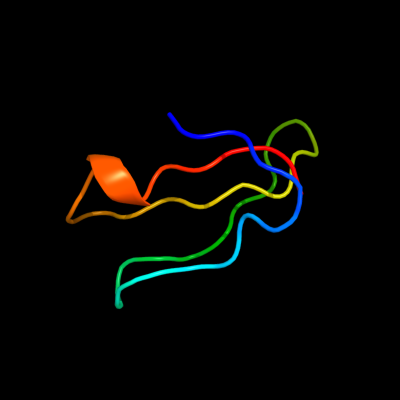
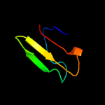
| 1 |
|
PDB 3bbo chain 6
Region: 1 - 38
Aligned: 38
Modelled: 38
Confidence: 99.9%
Identity: 71%
PDB header:ribosome
Chain: 6: PDB Molecule:ribosomal protein l36;
PDBTitle: homology model for the spinach chloroplast 50s subunit2 fitted to 9.4a cryo-em map of the 70s chlororibosome
Phyre2
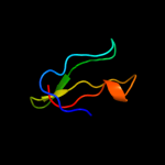
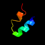
| 2 |
|
PDB 2qbk chain 4
Region: 1 - 38
Aligned: 38
Modelled: 38
Confidence: 99.9%
Identity: 100%
PDB header:ribosome
Chain: 4: PDB Molecule:50s ribosomal protein l36;
PDBTitle: crystal structure of the bacterial ribosome from escherichia2 coli in complex with gentamicin and ribosome recycling3 factor (rrf). this file contains the 50s subunit of the4 second 70s ribosome, with gentamicin and rrf bound. the5 entire crystal structure contains two 70s ribosomes and is6 described in remark 400.
Phyre2
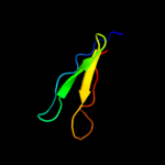
| 3 |
|
PDB 1dfe chain A
Region: 1 - 38
Aligned: 37
Modelled: 38
Confidence: 99.9%
Identity: 65%
Fold: Ribosomal protein L36
Superfamily: Ribosomal protein L36
Family: Ribosomal protein L36
Phyre2
| 4 |
|
PDB 2zjr chain 4 domain 1
Region: 1 - 38
Aligned: 37
Modelled: 38
Confidence: 99.9%
Identity: 68%
Fold: Ribosomal protein L36
Superfamily: Ribosomal protein L36
Family: Ribosomal protein L36
Phyre2
| 5 |
|
PDB 1j0t chain A
Region: 7 - 30
Aligned: 24
Modelled: 24
Confidence: 19.4%
Identity: 21%
Fold: Crustacean CHH/MIH/GIH neurohormone
Superfamily: Crustacean CHH/MIH/GIH neurohormone
Family: Crustacean CHH/MIH/GIH neurohormone
Phyre2
|
| Detailed template information | |
Due to computational demand, binding site predictions are not run for batch jobs
If you want to predict binding sites, please manually submit your model of choice to 3DLigandSite
Phyre is for academic use only
| Please cite: Protein structure prediction on
the web: a case study using the Phyre server |
| Kelley LA and Sternberg MJE. Nature Protocols
4, 363 - 371 (2009) [pdf] [Import into BibTeX] |
| |
| If you use the binding site
predictions from 3DLigandSite, please also cite: |
| 3DLigandSite: predicting ligand-binding sites using similar structures. |
| Wass MN, Kelley LA and Sternberg
MJ Nucleic Acids Research 38, W469-73 (2010) [PubMed] |
| |
|
|
|
|