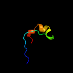
1 c3qr3B_
20.9
28
PDB header: hydrolaseChain: B: PDB Molecule: endoglucanase eg-ii;PDBTitle: crystal structure of cel5a (eg2) from hypocrea jecorina (trichoderma2 reesei)
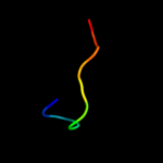
2 c3ifuA_
19.7
23
PDB header: transcriptionChain: A: PDB Molecule: non-structural protein;PDBTitle: the crystal structure of porcine reproductive and2 respiratory syndrome virus (prrsv) leader protease nsp1
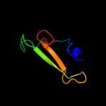
3 c2pnmA_
18.2
23
PDB header: hydrolaseChain: A: PDB Molecule: protease vp4;PDBTitle: crystal structure of vp4 protease from infectious pancreatic necrosis2 virus (ipnv) in space group p6122
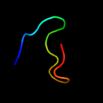
4 d2go8a1
12.4
33
Fold: Ferredoxin-likeSuperfamily: Dimeric alpha+beta barrelFamily: PG130-like5 d1yvua1
10.2
63
Fold: SH3-like barrelSuperfamily: PAZ domainFamily: PAZ domain6 c3bmzA_
9.0
39
PDB header: biosynthetic proteinChain: A: PDB Molecule: putative uncharacterized protein;PDBTitle: violacein biosynthetic enzyme vioe
7 d1lg4a_
8.7
21
Fold: Prion-likeSuperfamily: Prion-likeFamily: Prion-like8 d1rutx1
8.7
36
Fold: Glucocorticoid receptor-like (DNA-binding domain)Superfamily: Glucocorticoid receptor-like (DNA-binding domain)Family: LIM domain9 c1w62B_
8.2
17
PDB header: racemaseChain: B: PDB Molecule: b-cell mitogen;PDBTitle: proline racemase in complex with one molecule of pyrrole-2-2 carboxylic acid (hemi form)
10 d1i17a_
7.4
21
Fold: Prion-likeSuperfamily: Prion-likeFamily: Prion-like11 d1skza1
7.4
50
Fold: Knottins (small inhibitors, toxins, lectins)Superfamily: Leech antihemostatic proteinsFamily: Huristasin-like12 d1v47a1
7.4
29
Fold: PUA domain-likeSuperfamily: PUA domain-likeFamily: ATP sulfurylase N-terminal domain13 d1j2oa1
7.3
38
Fold: Glucocorticoid receptor-like (DNA-binding domain)Superfamily: Glucocorticoid receptor-like (DNA-binding domain)Family: LIM domain14 c2p0vA_
7.1
16
PDB header: structural genomics, unknown functionChain: A: PDB Molecule: hypothetical protein bt3781;PDBTitle: crystal structure of bt3781 protein from bacteroides2 thetaiotaomicron, northeast structural genomics target3 btr58
15 d2p0va1
7.1
16
Fold: alpha/alpha toroidSuperfamily: Six-hairpin glycosidasesFamily: CPF0428-like16 d1x6va1
7.0
29
Fold: PUA domain-likeSuperfamily: PUA domain-likeFamily: ATP sulfurylase N-terminal domain17 d1l5ja3
6.9
21
Fold: Aconitase iron-sulfur domainSuperfamily: Aconitase iron-sulfur domainFamily: Aconitase iron-sulfur domain18 c2jb1B_
6.9
24
PDB header: oxidoreductaseChain: B: PDB Molecule: l-amino acid oxidase;PDBTitle: the l-amino acid oxidase from rhodococcus opacus in complex2 with l-alanine
19 d1g8fa1
6.7
21
Fold: PUA domain-likeSuperfamily: PUA domain-likeFamily: ATP sulfurylase N-terminal domain20 d1jl0a_
6.7
33
Fold: S-adenosylmethionine decarboxylaseSuperfamily: S-adenosylmethionine decarboxylaseFamily: S-adenosylmethionine decarboxylase21 c2zkrg_
not modelled
6.6
36
PDB header: ribosomal protein/rnaChain: G: PDB Molecule: rna expansion segment es9;PDBTitle: structure of a mammalian ribosomal 60s subunit within an2 80s complex obtained by docking homology models of the rna3 and proteins into an 8.7 a cryo-em map
22 d1zata2
not modelled
6.2
0
Fold: L,D-transpeptidase pre-catalytic domain-likeSuperfamily: L,D-transpeptidase pre-catalytic domain-likeFamily: L,D-transpeptidase pre-catalytic domain-like23 d1m8pa1
not modelled
6.1
21
Fold: PUA domain-likeSuperfamily: PUA domain-likeFamily: ATP sulfurylase N-terminal domain24 c2hnhA_
not modelled
6.1
35
PDB header: transferaseChain: A: PDB Molecule: dna polymerase iii alpha subunit;PDBTitle: crystal structure of the catalytic alpha subunit of e. coli2 replicative dna polymerase iii
25 c2ezvA_
not modelled
6.0
18
PDB header: hydrolase/dnaChain: A: PDB Molecule: type ii restriction enzyme sfii;PDBTitle: crystal structure of tetrameric restriction endonuclease2 sfii bound to cognate dna.
26 d1u7ka_
not modelled
6.0
50
Fold: Retrovirus capsid protein, N-terminal core domainSuperfamily: Retrovirus capsid protein, N-terminal core domainFamily: Retrovirus capsid protein, N-terminal core domain27 d1yy9a4
not modelled
5.8
33
Fold: Knottins (small inhibitors, toxins, lectins)Superfamily: Growth factor receptor domainFamily: Growth factor receptor domain28 c2k2iB_
not modelled
5.6
36
PDB header: cell cycleChain: B: PDB Molecule: sfi1 peptide;PDBTitle: nmr solution structure of the c-terminal domain (t94-y172)2 of the human centrin 2 in complex with a repeat sequence of3 human sfi1 (r641-t660)
29 d1uzka3
not modelled
5.3
42
Fold: TB module/8-cys domainSuperfamily: TB module/8-cys domainFamily: TB module/8-cys domain30 d1jhda1
not modelled
5.2
21
Fold: PUA domain-likeSuperfamily: PUA domain-likeFamily: ATP sulfurylase N-terminal domain31 d1tm0a_
not modelled
5.2
15
Fold: Diaminopimelate epimerase-likeSuperfamily: Diaminopimelate epimerase-likeFamily: Proline racemase32 c3k4iC_
not modelled
5.0
31
PDB header: structural genomics, unknown functionChain: C: PDB Molecule: uncharacterized protein;PDBTitle: crystal structure of uncharacterized protein pspto_3204 from2 pseudomonas syringae pv. tomato str. dc3000