| Secondary structure and disorder prediction | |
| | |
1 | . | . | . | . | . | . | . | . | 10 | . | . | . | . | . | . | . | . | . | 20 | . | . | . | . | . | . | . | . | . | 30 | . | . | . | . | . | . | . | . | . | 40 | . | . | . | . | . | . | . | . | . | 50 | . | . | . | . | . | . | . | . | . | 60 |
| Sequence | |
M | M | K | R | V | S | M | N | E | W | I | M | P | V | L | I | Y | A | Q | N | T | M | L | N | L | K | L | T | I | P | G | L | R | A | E | R | K | V | I | S | K | L | S | P | R | S | Y | L | K | R | S | L | I | P | I | K | K | I | E | T |
| Secondary structure | |
|
|  | | |  |
|
|
| | | | | | | | | | |
| | | | | | | | | |
|
|
|  | | | | | | | |
|
|
| | | | | | | | |
|
|
| | | | | |
|
| SS confidence | |
|
|
|
|
|
|
|
|
|
|
|
|
|
|
|
|
|
|
|
|
|
|
|
|
|
|
|
|
|
|
|
|
|
|
|
|
|
|
|
|
|
|
|
|
|
|
|
|
|
|
|
|
|
|
|
|
|
|
|
|
| Disorder | |
? | ? | ? | ? | ? | ? | ? |
|
|
|
|
|
|
|
|
|
|
|
|
|
|
|
|
|
|
|
|
|
| ? |
|
|
| ? |
|
|
|
|
|
|
|
|
| ? |
|
|
|
|
|
|
|
|
|
|
|
| ? | ? |
| ? |
| Disorder confidence | |
|
|
|
|
|
|
|
|
|
|
|
|
|
|
|
|
|
|
|
|
|
|
|
|
|
|
|
|
|
|
|
|
|
|
|
|
|
|
|
|
|
|
|
|
|
|
|
|
|
|
|
|
|
|
|
|
|
|
|
|
| |
| | |
. | . | . | . | . | . | . | . | . | 70 | . | . | . | . | . |
| Sequence | |
K | I | D | S | V | E | C | I | S | I | H | R | V | D | S |
| Secondary structure | |
|
|
| | | | | | | | | | | |
|
| SS confidence | |
|
|
|
|
|
|
|
|
|
|
|
|
|
|
|
| Disorder | |
|
|
|
|
|
|
|
|
|
|
| ? | ? | ? | ? |
| Disorder confidence | |
|
|
|
|
|
|
|
|
|
|
|
|
|
|
|
| |
| Confidence Key |
| High(9) | |
|
|
|
|
|
|
|
|
|
Low (0) |
| ? | Disordered |
| Alpha helix |
| Beta strand |
Hover over an aligned region to see model and summary info
Please note, only up to the top 20 hits are modelled to reduce computer load
|
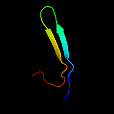
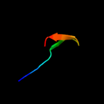
| 1 |
|
PDB 3gla chain A
Region: 9 - 35
Aligned: 27
Modelled: 27
Confidence: 20.4%
Identity: 22%
PDB header:chaperone
Chain: A: PDB Molecule:low molecular weight heat shock protein;
PDBTitle: crystal structure of the hspa from xanthomonas axonopodis
Phyre2
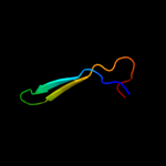
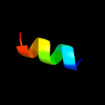
| 2 |
|
PDB 1bdf chain A domain 1
Region: 6 - 66
Aligned: 61
Modelled: 61
Confidence: 13.7%
Identity: 21%
Fold: DCoH-like
Superfamily: RBP11-like subunits of RNA polymerase
Family: RNA polymerase alpha subunit dimerisation domain
Phyre2
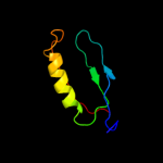
| 3 |
|
PDB 2kcd chain A
Region: 11 - 25
Aligned: 15
Modelled: 15
Confidence: 12.7%
Identity: 27%
PDB header:structural genomics, unknown function
Chain: A: PDB Molecule:uncharacterized protein ssp0047;
PDBTitle: solution nmr structure of ssp0047 from staphylococcus2 saprophyticus. northeast structural genomics consortium3 target syr6.
Phyre2
| 4 |
|
PDB 2pq4 chain B
Region: 41 - 49
Aligned: 9
Modelled: 9
Confidence: 6.6%
Identity: 67%
PDB header:chaperone/oxidoreductase
Chain: B: PDB Molecule:periplasmic nitrate reductase precursor;
PDBTitle: nmr solution structure of napd in complex with napa1-352 signal peptide
Phyre2
| 5 |
|
PDB 1avy chain A
Region: 55 - 65
Aligned: 11
Modelled: 11
Confidence: 6.2%
Identity: 73%
PDB header:coiled coil
Chain: A: PDB Molecule:fibritin;
PDBTitle: fibritin deletion mutant m (bacteriophage t4)
Phyre2
|
| Detailed template information | |
Due to computational demand, binding site predictions are not run for batch jobs
If you want to predict binding sites, please manually submit your model of choice to 3DLigandSite
Phyre is for academic use only
| Please cite: Protein structure prediction on
the web: a case study using the Phyre server |
| Kelley LA and Sternberg MJE. Nature Protocols
4, 363 - 371 (2009) [pdf] [Import into BibTeX] |
| |
| If you use the binding site
predictions from 3DLigandSite, please also cite: |
| 3DLigandSite: predicting ligand-binding sites using similar structures. |
| Wass MN, Kelley LA and Sternberg
MJ Nucleic Acids Research 38, W469-73 (2010) [PubMed] |
| |
|
|
|
|