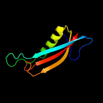
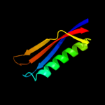
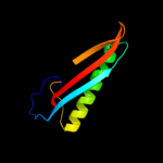
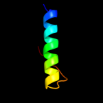
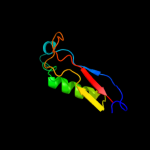
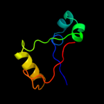
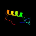
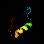
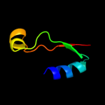
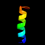
PDB header:membrane protein Chain: A: PDB Molecule:putative outer membrane protein, signal; PDBTitle: crystal structure of the e. coli outer membrane lipoprotein2 rcsf
Confidence and coverage
Confidence:
97.9%
Coverage:
68%
80 residues ( 68% of your sequence) have been modelled with 97.9% confidence by the single highest scoring template.
You may wish to submit your sequence to Phyrealarm. This will automatically scan your sequence every week for new potential templates as they appear in the Phyre2 library.
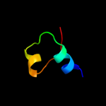
Region: 34 - 112 Aligned: 78 Modelled: 79 Confidence: 95.3% Identity: 15% PDB header:structural genomics, unknown function Chain: B: PDB Molecule:uncharacterized protein; PDBTitle: crystal structure of a protein with unknown function which belongs to2 pfam duf74 family (pepe_0654) from pediococcus pentosaceus atcc 257453 at 2.73 a resolution
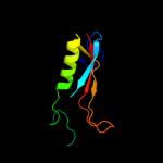
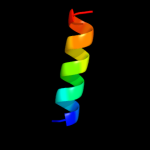
Region: 63 - 91 Aligned: 29 Modelled: 29 Confidence: 34.6% Identity: 7% PDB header:ligase Chain: A: PDB Molecule:threonyl-trna synthetase; PDBTitle: crystal structure of the editing domain of threonyl-trna2 synthetase from pyrococcus abyssi in complex with an3 analog of seryladenylate
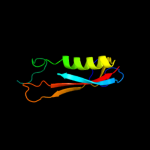
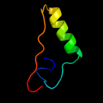
Region: 66 - 85 Aligned: 20 Modelled: 20 Confidence: 8.6% Identity: 30% PDB header:protein binding Chain: B: PDB Molecule:peptide of far upstream element-binding protein 1; PDBTitle: solution structure of the first two rrm domains of fir in the complex2 with fbp nbox peptide