| 1 |
|
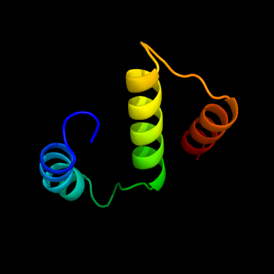
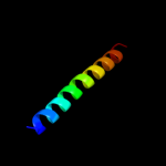
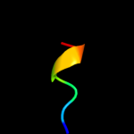
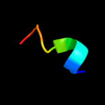
PDB 2i68 chain B
Region: 22 - 98
Aligned: 58
Modelled: 59
Confidence: 24.2%
Identity: 19%
PDB header:transport protein
Chain: B: PDB Molecule:protein emre;
PDBTitle: cryo-em based theoretical model structure of transmembrane2 domain of the multidrug-resistance antiporter from e. coli3 emre
Phyre2
| 2 |
|
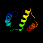
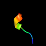
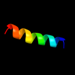
PDB 1xoo chain A
Region: 24 - 33
Aligned: 10
Modelled: 10
Confidence: 11.9%
Identity: 70%
PDB header:viral protein
Chain: A: PDB Molecule:hemagglutinin;
PDBTitle: nmr structure of g1s mutant of influenza hemagglutinin2 fusion peptide in dpc micelles at ph 5
Phyre2
| 3 |
|
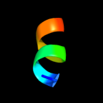
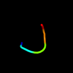
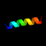
PDB 2y69 chain W
Region: 72 - 102
Aligned: 31
Modelled: 31
Confidence: 10.5%
Identity: 19%
PDB header:electron transport
Chain: W: PDB Molecule:cytochrome c oxidase polypeptide 7a1;
PDBTitle: bovine heart cytochrome c oxidase re-refined with molecular2 oxygen
Phyre2
| 4 |
|
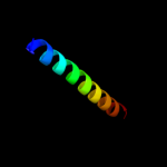
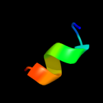
PDB 1v54 chain J
Region: 72 - 102
Aligned: 31
Modelled: 31
Confidence: 10.3%
Identity: 19%
Fold: Single transmembrane helix
Superfamily: Mitochondrial cytochrome c oxidase subunit VIIa
Family: Mitochondrial cytochrome c oxidase subunit VIIa
Phyre2
| 5 |
|
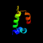
PDB 1htj chain F
Region: 11 - 57
Aligned: 41
Modelled: 47
Confidence: 10.2%
Identity: 22%
Fold: Regulator of G-protein signaling, RGS
Superfamily: Regulator of G-protein signaling, RGS
Family: Regulator of G-protein signaling, RGS
Phyre2
| 6 |
|
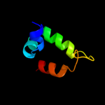
PDB 1htj chain F
Region: 11 - 57
Aligned: 41
Modelled: 47
Confidence: 10.2%
Identity: 22%
PDB header:signaling protein
Chain: F: PDB Molecule:kiaa0380;
PDBTitle: structure of the rgs-like domain from pdz-rhogef
Phyre2
| 7 |
|
PDB 2kxa chain A
Region: 27 - 33
Aligned: 7
Modelled: 7
Confidence: 7.5%
Identity: 71%
PDB header:viral protein, immune system
Chain: A: PDB Molecule:haemagglutinin ha2 chain peptide;
PDBTitle: the hemagglutinin fusion peptide (h1 subtype) at ph 7.4
Phyre2
| 8 |
|
PDB 1xop chain A
Region: 27 - 33
Aligned: 7
Modelled: 7
Confidence: 6.7%
Identity: 71%
PDB header:viral protein
Chain: A: PDB Molecule:hemagglutinin;
PDBTitle: nmr structure of g1v mutant of influenza hemagglutinin2 fusion peptide in dpc micelles at ph 5
Phyre2
| 9 |
|
PDB 1ibo chain A
Region: 27 - 33
Aligned: 7
Modelled: 7
Confidence: 6.7%
Identity: 71%
PDB header:viral protein
Chain: A: PDB Molecule:hemagglutinin ha2 chain peptide;
PDBTitle: nmr structure of hemagglutinin fusion peptide in dpc2 micelles at ph 7.4
Phyre2
| 10 |
|
PDB 1ibn chain A
Region: 27 - 33
Aligned: 7
Modelled: 7
Confidence: 6.7%
Identity: 71%
PDB header:viral protein
Chain: A: PDB Molecule:hemagglutinin ha2 chain peptide;
PDBTitle: nmr structure of hemagglutinin fusion peptide in dpc2 micelles at ph 5
Phyre2
| 11 |
|
PDB 2kn8 chain A
Region: 58 - 62
Aligned: 5
Modelled: 5
Confidence: 6.7%
Identity: 60%
PDB header:protein binding, dna binding protein
Chain: A: PDB Molecule:dna cleavage and packaging protein large subunit, ul89;
PDBTitle: nmr structure of the c-terminal domain of pul89
Phyre2
| 12 |
|
PDB 2l4g chain A
Region: 25 - 33
Aligned: 9
Modelled: 9
Confidence: 6.5%
Identity: 67%
PDB header:viral protein
Chain: A: PDB Molecule:haemagglutinin;
PDBTitle: influenza haemagglutinin fusion peptide mutant g13a
Phyre2
| 13 |
|
PDB 2dci chain A
Region: 25 - 33
Aligned: 9
Modelled: 9
Confidence: 6.2%
Identity: 67%
PDB header:viral protein
Chain: A: PDB Molecule:hemagglutinin;
PDBTitle: nmr structure of influenza ha fusion peptide mutant w14a in2 dpc in ph5
Phyre2
| 14 |
|
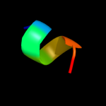
PDB 3a0h chain X
Region: 60 - 78
Aligned: 19
Modelled: 19
Confidence: 5.1%
Identity: 42%
PDB header:electron transport
Chain: X: PDB Molecule:photosystem ii reaction center protein x;
PDBTitle: crystal structure of i-substituted photosystem ii complex
Phyre2
| 15 |
|
PDB 3a0b chain X
Region: 60 - 78
Aligned: 19
Modelled: 19
Confidence: 5.1%
Identity: 42%
PDB header:electron transport
Chain: X: PDB Molecule:photosystem ii reaction center protein x;
PDBTitle: crystal structure of br-substituted photosystem ii complex
Phyre2
| 16 |
|
PDB 3a0b chain X
Region: 60 - 78
Aligned: 19
Modelled: 19
Confidence: 5.1%
Identity: 42%
PDB header:electron transport
Chain: X: PDB Molecule:photosystem ii reaction center protein x;
PDBTitle: crystal structure of br-substituted photosystem ii complex
Phyre2