| 1 |
|
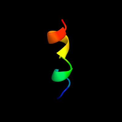
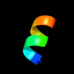
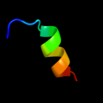
PDB 1emz chain A
Region: 18 - 30
Aligned: 13
Modelled: 13
Confidence: 14.3%
Identity: 62%
PDB header:viral protein
Chain: A: PDB Molecule:envelope glycoprotein e1;
PDBTitle: solution structure of fragment (350-370) of the2 transmembrane domain of hepatitis c envelope glycoprotein3 e1
Phyre2
| 2 |
|
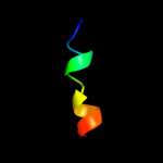
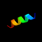
PDB 2hg5 chain D
Region: 18 - 35
Aligned: 18
Modelled: 18
Confidence: 13.1%
Identity: 22%
PDB header:membrane protein
Chain: D: PDB Molecule:kcsa channel;
PDBTitle: cs+ complex of a k channel with an amide to ester substitution in the2 selectivity filter
Phyre2
| 3 |
|
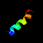
PDB 2fok chain A domain 2
Region: 24 - 33
Aligned: 10
Modelled: 10
Confidence: 7.1%
Identity: 60%
Fold: DNA/RNA-binding 3-helical bundle
Superfamily: "Winged helix" DNA-binding domain
Family: Restriction endonuclease FokI, N-terminal (recognition) domain
Phyre2
| 4 |
|
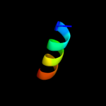
PDB 3qfh chain E
Region: 14 - 27
Aligned: 14
Modelled: 14
Confidence: 7.1%
Identity: 50%
PDB header:hydrolase
Chain: E: PDB Molecule:epidermin leader peptide processing serine protease epip;
PDBTitle: 2.05 angstrom resolution crystal structure of epidermin leader peptide2 processing serine protease (epip) from staphylococcus aureus.
Phyre2
| 5 |
|
PDB 1kve chain A
Region: 16 - 32
Aligned: 17
Modelled: 17
Confidence: 6.9%
Identity: 47%
PDB header:toxin
Chain: A: PDB Molecule:smk toxin;
PDBTitle: killer toxin from halotolerant yeast
Phyre2
| 6 |
|
PDB 3a9f chain A
Region: 24 - 34
Aligned: 11
Modelled: 11
Confidence: 6.7%
Identity: 36%
PDB header:electron transport
Chain: A: PDB Molecule:cytochrome c;
PDBTitle: crystal structure of the c-terminal domain of cytochrome cz2 from chlorobium tepidum
Phyre2
| 7 |
|
PDB 2b6n chain A
Region: 14 - 27
Aligned: 14
Modelled: 14
Confidence: 6.4%
Identity: 36%
PDB header:hydrolase
Chain: A: PDB Molecule:proteinase k;
PDBTitle: the 1.8 a crystal structure of a proteinase k like enzyme from a2 psychrotroph serratia species
Phyre2
| 8 |
|
PDB 2b0l chain A domain 1
Region: 20 - 33
Aligned: 14
Modelled: 14
Confidence: 6.3%
Identity: 57%
Fold: DNA/RNA-binding 3-helical bundle
Superfamily: "Winged helix" DNA-binding domain
Family: CodY HTH domain
Phyre2