Phyre Home Retrieve Phyre Job Id
Emaill.a.kelley@imperial.ac.uk
DescriptionP0AAU5
DateThu Jan 5 11:13:51 GMT 2012
Unique Job IDe9f411aeb376f30a

Summary 

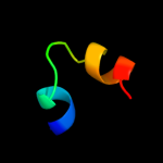
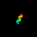
Top model
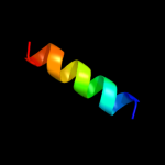
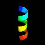
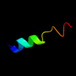
Image coloured by rainbow N → C terminus
Model (left) based on template c2hg5D_
Top template information
PDB header:membrane protein
Chain: D: PDB Molecule:kcsa channel;
PDBTitle: cs+ complex of a k channel with an amide to ester substitution in the2 selectivity filter
Confidence and coverage
Confidence: 26.3% Coverage: 15%
16 residues ( 15% of your sequence) have been modelled with 26.3% confidence by the single highest scoring template.
You may wish to submit your sequence to Phyrealarm. This will automatically scan your sequence every week for new potential templates as they appear in the Phyre2 library.
Please note: You must be registered and logged in to use Phyrealarm.
3D viewing
Interactive 3D view in Jmol

Sequence analysis 

Secondary structure and disorder prediction 

   1........10.........20.........30.........40.........50.........60
Sequence  MKYIIFLFRAIWLALSLLILFFSMHRLSLLDSTRDVSELISLMSYGMMVICFPTGIVFFI
Secondary structure 


SS confidence 


Disorder  ?


Disorder confidence 


   .........70.........80.........90.........100........
Sequence  ALIFIGTVSDIIGVRIDSKYIMAIIIWLYFLSGGYIQWFVLSKRIINK
Secondary structure 

SS confidence 


Disorder 


?


???
Disorder confidence 


Confidence Key
High(9)                    Low (0)
?Disordered
Alpha helix
Beta strand

Domain analysis 

Hover over an aligned region to see model and summary info

Please note, only up to the top 20 hits are modelled to reduce computer load

RankAligned region

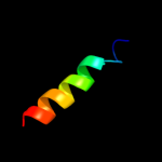
PDB 2hg5 chain D

3D model

Region: 42 - 57
Aligned: 16
Modelled: 16
Confidence: 26.3%
Identity: 19%
PDB header:membrane protein
Chain: D: PDB Molecule:kcsa channel;
PDBTitle: cs+ complex of a k channel with an amide to ester substitution in the2 selectivity filter

Phyre2

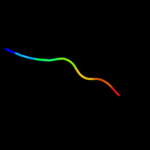
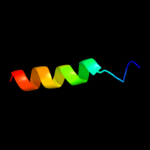
PDB 2krx chain A

3D model

Region: 93 - 99
Aligned: 7
Modelled: 7
Confidence: 26.1%
Identity: 57%
PDB header:structural genomics, unknown function
Chain: A: PDB Molecule:asl3597 protein;
PDBTitle: solution nmr structure of asl3597 from nostoc sp. pcc7120.2 northeast structural genomics consortium target id nsr244.

Phyre2

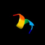
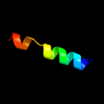
PDB 1w8x chain P

3D model

Region: 84 - 90
Aligned: 7
Modelled: 7
Confidence: 21.1%
Identity: 71%
PDB header:virus
Chain: P: PDB Molecule:protein p16;
PDBTitle: structural analysis of prd1

Phyre2

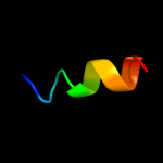
PDB 1rkc chain B

3D model

Region: 29 - 41
Aligned: 13
Modelled: 13
Confidence: 17.7%
Identity: 54%
PDB header:cell adhesion, structural protein
Chain: B: PDB Molecule:talin;
PDBTitle: human vinculin head (1-258) in complex with talin's2 vinculin binding site 3 (residues 1944-1969)

Phyre2

PDB 1xwj chain B

3D model

Region: 29 - 41
Aligned: 13
Modelled: 13
Confidence: 17.4%
Identity: 54%
PDB header:cell adhesion/protein binding
Chain: B: PDB Molecule:talin;
PDBTitle: vinculin head (1-258) in complex with the talin vinculin2 binding site 3 (1945-1969)

Phyre2

PDB 1xme chain B domain 2

3D model

Region: 5 - 21
Aligned: 17
Modelled: 17
Confidence: 15.7%
Identity: 47%
Fold: Transmembrane helix hairpin
Superfamily: Cytochrome c oxidase subunit II-like, transmembrane region
Family: Cytochrome c oxidase subunit II-like, transmembrane region

Phyre2

PDB 1smy chain E

3D model

Region: 70 - 86
Aligned: 17
Modelled: 17
Confidence: 14.2%
Identity: 29%
Fold: RPB6/omega subunit-like
Superfamily: RPB6/omega subunit-like
Family: RNA polymerase omega subunit

Phyre2

PDB 2gyc chain 3 domain 1

3D model

Region: 32 - 43
Aligned: 12
Modelled: 12
Confidence: 11.5%
Identity: 67%
Fold: Ribosomal protein L7/12, oligomerisation (N-terminal) domain
Superfamily: Ribosomal protein L7/12, oligomerisation (N-terminal) domain
Family: Ribosomal protein L7/12, oligomerisation (N-terminal) domain

Phyre2

PDB 1ynj chain K domain 1

3D model

Region: 70 - 86
Aligned: 17
Modelled: 17
Confidence: 10.8%
Identity: 29%
Fold: RPB6/omega subunit-like
Superfamily: RPB6/omega subunit-like
Family: RNA polymerase omega subunit

Phyre2

PDB 2a5y chain B domain 2

3D model

Region: 33 - 44
Aligned: 12
Modelled: 12
Confidence: 9.5%
Identity: 75%
Fold: DEATH domain
Superfamily: DEATH domain
Family: Caspase recruitment domain, CARD

Phyre2

PDB 1vkn chain A domain 2

3D model

Region: 51 - 61
Aligned: 11
Modelled: 11
Confidence: 9.5%
Identity: 36%
Fold: FwdE/GAPDH domain-like
Superfamily: Glyceraldehyde-3-phosphate dehydrogenase-like, C-terminal domain
Family: GAPDH-like

Phyre2

PDB 1rqt chain A

3D model

Region: 32 - 43
Aligned: 12
Modelled: 12
Confidence: 9.2%
Identity: 67%
PDB header:ribosome
Chain: A: PDB Molecule:50s ribosomal protein l7/l12;
PDBTitle: nmr structure of dimeric n-terminal domain of ribosomal2 protein l7 from e.coli

Phyre2

PDB 1rqt chain A

3D model

Region: 32 - 43
Aligned: 12
Modelled: 12
Confidence: 9.2%
Identity: 67%
Fold: Ribosomal protein L7/12, oligomerisation (N-terminal) domain
Superfamily: Ribosomal protein L7/12, oligomerisation (N-terminal) domain
Family: Ribosomal protein L7/12, oligomerisation (N-terminal) domain

Phyre2

PDB 1rqt chain B

3D model

Region: 32 - 43
Aligned: 12
Modelled: 12
Confidence: 9.2%
Identity: 67%
PDB header:ribosome
Chain: B: PDB Molecule:50s ribosomal protein l7/l12;
PDBTitle: nmr structure of dimeric n-terminal domain of ribosomal2 protein l7 from e.coli

Phyre2

PDB 2e74 chain E domain 1

3D model

Region: 55 - 73
Aligned: 19
Modelled: 19
Confidence: 8.8%
Identity: 47%
Fold: Single transmembrane helix
Superfamily: PetL subunit of the cytochrome b6f complex
Family: PetL subunit of the cytochrome b6f complex

Phyre2

PDB 2e74 chain E

3D model

Region: 55 - 73
Aligned: 19
Modelled: 19
Confidence: 8.8%
Identity: 47%
PDB header:photosynthesis
Chain: E: PDB Molecule:cytochrome b6-f complex subunit 6;
PDBTitle: crystal structure of the cytochrome b6f complex from m.laminosus

Phyre2

PDB 2e75 chain E

3D model

Region: 55 - 73
Aligned: 19
Modelled: 19
Confidence: 8.8%
Identity: 47%
PDB header:photosynthesis
Chain: E: PDB Molecule:cytochrome b6-f complex subunit 6;
PDBTitle: crystal structure of the cytochrome b6f complex with 2-nonyl-4-2 hydroxyquinoline n-oxide (nqno) from m.laminosus

Phyre2

PDB 1vf5 chain R

3D model

Region: 55 - 73
Aligned: 19
Modelled: 19
Confidence: 8.8%
Identity: 47%
PDB header:photosynthesis
Chain: R: PDB Molecule:protein pet l;
PDBTitle: crystal structure of cytochrome b6f complex from m.laminosus

Phyre2

PDB 1vf5 chain E

3D model

Region: 55 - 73
Aligned: 19
Modelled: 19
Confidence: 8.8%
Identity: 47%
PDB header:photosynthesis
Chain: E: PDB Molecule:protein pet l;
PDBTitle: crystal structure of cytochrome b6f complex from m.laminosus

Phyre2

PDB 2e76 chain E

3D model

Region: 55 - 73
Aligned: 19
Modelled: 19
Confidence: 8.8%
Identity: 47%
PDB header:photosynthesis
Chain: E: PDB Molecule:cytochrome b6-f complex subunit 6;
PDBTitle: crystal structure of the cytochrome b6f complex with tridecyl-2 stigmatellin (tds) from m.laminosus

Phyre2
1

c2hg5D_
2

c2krxA_
3

c1w8xP_
4

c1rkcB_
5

c1xwjB_
6

d1xmeb2
7

d1smye_
8

d2gyc31
9

d1ynjk1
10

d2a5yb2
11

d1vkna2
12

c1rqtA_
13

d1rqta_
14

c1rqtB_
15

d2e74e1
16

c2e74E_
17

c2e75E_
18

c1vf5R_
19

c1vf5E_
20

c2e76E_
21


22


23


24


25


26


27


28


29


30


31


32


33


34


35


Detailed template information 

#
Template Alignment Coverage3D Model Confidence
% i.d. Template Information
1c2hg5D_


26.3 19 PDB header:membrane protein
Chain: D: PDB Molecule:kcsa channel;
PDBTitle: cs+ complex of a k channel with an amide to ester substitution in the2 selectivity filter
2c2krxA_


26.1 57 PDB header:structural genomics, unknown function
Chain: A: PDB Molecule:asl3597 protein;
PDBTitle: solution nmr structure of asl3597 from nostoc sp. pcc7120.2 northeast structural genomics consortium target id nsr244.
3c1w8xP_


21.1 71 PDB header:virus
Chain: P: PDB Molecule:protein p16;
PDBTitle: structural analysis of prd1
4c1rkcB_


17.7 54 PDB header:cell adhesion, structural protein
Chain: B: PDB Molecule:talin;
PDBTitle: human vinculin head (1-258) in complex with talin's2 vinculin binding site 3 (residues 1944-1969)
5c1xwjB_


17.4 54 PDB header:cell adhesion/protein binding
Chain: B: PDB Molecule:talin;
PDBTitle: vinculin head (1-258) in complex with the talin vinculin2 binding site 3 (1945-1969)
6d1xmeb2


15.7 47 Fold:Transmembrane helix hairpin
Superfamily:Cytochrome c oxidase subunit II-like, transmembrane region
Family:Cytochrome c oxidase subunit II-like, transmembrane region
7d1smye_


14.2 29 Fold:RPB6/omega subunit-like
Superfamily:RPB6/omega subunit-like
Family:RNA polymerase omega subunit
8d2gyc31


11.5 67 Fold:Ribosomal protein L7/12, oligomerisation (N-terminal) domain
Superfamily:Ribosomal protein L7/12, oligomerisation (N-terminal) domain
Family:Ribosomal protein L7/12, oligomerisation (N-terminal) domain
9d1ynjk1


10.8 29 Fold:RPB6/omega subunit-like
Superfamily:RPB6/omega subunit-like
Family:RNA polymerase omega subunit
10d2a5yb2


9.5 75 Fold:DEATH domain
Superfamily:DEATH domain
Family:Caspase recruitment domain, CARD
11d1vkna2


9.5 36 Fold:FwdE/GAPDH domain-like
Superfamily:Glyceraldehyde-3-phosphate dehydrogenase-like, C-terminal domain
Family:GAPDH-like
12c1rqtA_


9.2 67 PDB header:ribosome
Chain: A: PDB Molecule:50s ribosomal protein l7/l12;
PDBTitle: nmr structure of dimeric n-terminal domain of ribosomal2 protein l7 from e.coli
13d1rqta_


9.2 67 Fold:Ribosomal protein L7/12, oligomerisation (N-terminal) domain
Superfamily:Ribosomal protein L7/12, oligomerisation (N-terminal) domain
Family:Ribosomal protein L7/12, oligomerisation (N-terminal) domain
14c1rqtB_


9.2 67 PDB header:ribosome
Chain: B: PDB Molecule:50s ribosomal protein l7/l12;
PDBTitle: nmr structure of dimeric n-terminal domain of ribosomal2 protein l7 from e.coli
15d2e74e1


8.8 47 Fold:Single transmembrane helix
Superfamily:PetL subunit of the cytochrome b6f complex
Family:PetL subunit of the cytochrome b6f complex
16c2e74E_


8.8 47 PDB header:photosynthesis
Chain: E: PDB Molecule:cytochrome b6-f complex subunit 6;
PDBTitle: crystal structure of the cytochrome b6f complex from m.laminosus
17c2e75E_


8.8 47 PDB header:photosynthesis
Chain: E: PDB Molecule:cytochrome b6-f complex subunit 6;
PDBTitle: crystal structure of the cytochrome b6f complex with 2-nonyl-4-2 hydroxyquinoline n-oxide (nqno) from m.laminosus
18c1vf5R_


8.8 47 PDB header:photosynthesis
Chain: R: PDB Molecule:protein pet l;
PDBTitle: crystal structure of cytochrome b6f complex from m.laminosus
19c1vf5E_


8.8 47 PDB header:photosynthesis
Chain: E: PDB Molecule:protein pet l;
PDBTitle: crystal structure of cytochrome b6f complex from m.laminosus
20c2e76E_


8.8 47 PDB header:photosynthesis
Chain: E: PDB Molecule:cytochrome b6-f complex subunit 6;
PDBTitle: crystal structure of the cytochrome b6f complex with tridecyl-2 stigmatellin (tds) from m.laminosus
21d2q49a2


not modelled 8.7 29 Fold:FwdE/GAPDH domain-like
Superfamily:Glyceraldehyde-3-phosphate dehydrogenase-like, C-terminal domain
Family:GAPDH-like
22d2g17a2


not modelled 8.2 27 Fold:FwdE/GAPDH domain-like
Superfamily:Glyceraldehyde-3-phosphate dehydrogenase-like, C-terminal domain
Family:GAPDH-like
23d1whba_


not modelled 7.8 50 Fold:Rhodanese/Cell cycle control phosphatase
Superfamily:Rhodanese/Cell cycle control phosphatase
Family:Ubiquitin carboxyl-terminal hydrolase 8, USP8
24c3jycA_


not modelled 7.5 21 PDB header:metal transport
Chain: A: PDB Molecule:inward-rectifier k+ channel kir2.2;
PDBTitle: crystal structure of the eukaryotic strong inward-rectifier2 k+ channel kir2.2 at 3.1 angstrom resolution
25c2kpeB_


not modelled 7.5 33 PDB header:membrane protein
Chain: B: PDB Molecule:glycophorin-a;
PDBTitle: refined structure of glycophorin a transmembrane segment dimer in dpc2 micelles
26c2kpeA_


not modelled 7.5 33 PDB header:membrane protein
Chain: A: PDB Molecule:glycophorin-a;
PDBTitle: refined structure of glycophorin a transmembrane segment dimer in dpc2 micelles
27d2gwfa1


not modelled 7.2 50 Fold:Rhodanese/Cell cycle control phosphatase
Superfamily:Rhodanese/Cell cycle control phosphatase
Family:Ubiquitin carboxyl-terminal hydrolase 8, USP8
28d1ymka1


not modelled 6.6 55 Fold:Rhodanese/Cell cycle control phosphatase
Superfamily:Rhodanese/Cell cycle control phosphatase
Family:Cell cycle control phosphatase, catalytic domain
29c2ojlB_


not modelled 6.6 50 PDB header:structural genomics, unknown function
Chain: B: PDB Molecule:hypothetical protein;
PDBTitle: crystal structure of q7waf1_borpa from bordetella parapertussis.2 northeast structural genomics target bpr68.
30c2p0gB_


not modelled 6.5 38 PDB header:structural genomics, unknown function
Chain: B: PDB Molecule:selenoprotein w-related protein;
PDBTitle: crystal structure of selenoprotein w-related protein from2 vibrio cholerae. northeast structural genomics target vcr75
31d1pgya_


not modelled 6.4 47 Fold:RuvA C-terminal domain-like
Superfamily:UBA-like
Family:UBA domain
32d1pd0a4


not modelled 6.1 27 Fold:Gelsolin-like
Superfamily:C-terminal, gelsolin-like domain of Sec23/24
Family:C-terminal, gelsolin-like domain of Sec23/24
33c2l0oA_


not modelled 6.0 50 PDB header:membrane protein
Chain: A: PDB Molecule:oxidoreductase that catalyzes reoxidation of dsba protein
PDBTitle: dsbb3 peptide structure in 100% tfe
34d2fa8a1


not modelled 5.8 44 Fold:Thioredoxin fold
Superfamily:Thioredoxin-like
Family:Selenoprotein W-related
35c3dexA_


not modelled 5.5 56 PDB header:structural genomics, unknown function
Chain: A: PDB Molecule:sav_2001;
PDBTitle: crystal structure of sav_2001 protein from streptomyces2 avermitilis, northeast structural genomics consortium3 target svr107.

Binding site prediction 

Due to computational demand, binding site predictions are not run for batch jobs

If you want to predict binding sites, please manually submit your model of choice to 3DLigandSite


Phyre is for academic use only

Please cite: Protein structure prediction on the web: a case study using the Phyre server
Kelley LA and Sternberg MJE. Nature Protocols 4, 363 - 371 (2009) [pdf] [Import into BibTeX]
 
If you use the binding site predictions from 3DLigandSite, please also cite:
3DLigandSite: predicting ligand-binding sites using similar structures.
Wass MN, Kelley LA and Sternberg MJ Nucleic Acids Research 38, W469-73 (2010) [PubMed]
 
© Structural Bioinformatics Group
Imperial College London
Lawrence Kelley, Benjamin Jefferys
Disclaimer
Terms and Conditions
Component software
Template detection: HHpred 1.51
Secondary structure prediction: Psi-pred 2.5
Disorder prediction: Disopred 2.4
Transmembrane prediction: Memsat_SVM
Multi-template modelling and ab initio: Poing 1.0