Phyre Home Retrieve Phyre Job Id
Emaill.a.kelley@imperial.ac.uk
DescriptionP67624
DateThu Jan 5 12:10:44 GMT 2012
Unique Job IDe5a3907fea9a7a6f

Summary 

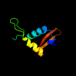
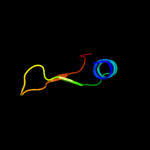
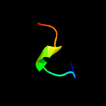
Top model
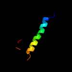
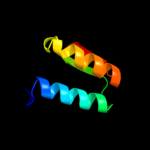
Image coloured by rainbow N → C terminus
Model (left) based on template d1y0na_
Top template information
Fold:YehU-like
Superfamily:YehU-like
Family:YehU-like
Confidence and coverage
Confidence:100.0% Coverage: 86%
62 residues ( 86% of your sequence) have been modelled with 100.0% confidence by the single highest scoring template.
3D viewing
Interactive 3D view in Jmol

Sequence analysis 

Secondary structure and disorder prediction 

   1........10.........20.........30.........40.........50.........60
Sequence  MLIPWQDLSPETLENLIESFVLREGTDYGEHERTLEQKVADVKRQLQCGEAVLVWSELHE
Secondary structure 


SS confidence 


Disorder  ?????


Disorder confidence 


   .........70..
Sequence  TVNIMPRSQFRE
Secondary structure 

SS confidence 


Disorder 


?


???
Disorder confidence 


Confidence Key
High(9)                    Low (0)
?Disordered
Alpha helix
Beta strand

Domain analysis 

Hover over an aligned region to see model and summary info

Please note, only up to the top 20 hits are modelled to reduce computer load

RankAligned region

PDB 1y0n chain A

3D model

Region: 1 - 70
Aligned: 62
Modelled: 70
Confidence: 100.0%
Identity: 39%
Fold: YehU-like
Superfamily: YehU-like
Family: YehU-like

Phyre2

PDB 1g57 chain A

3D model

Region: 37 - 68
Aligned: 31
Modelled: 32
Confidence: 83.2%
Identity: 26%
Fold: YrdC/RibB
Superfamily: YrdC/RibB
Family: 3,4-dihydroxy-2-butanone 4-phosphate synthase, DHBP synthase, RibB

Phyre2

PDB 1snn chain A

3D model

Region: 38 - 68
Aligned: 31
Modelled: 31
Confidence: 74.3%
Identity: 26%
Fold: YrdC/RibB
Superfamily: YrdC/RibB
Family: 3,4-dihydroxy-2-butanone 4-phosphate synthase, DHBP synthase, RibB

Phyre2

PDB 1k4i chain A

3D model

Region: 39 - 68
Aligned: 30
Modelled: 30
Confidence: 68.4%
Identity: 27%
Fold: YrdC/RibB
Superfamily: YrdC/RibB
Family: 3,4-dihydroxy-2-butanone 4-phosphate synthase, DHBP synthase, RibB

Phyre2

PDB 3mio chain A

3D model

Region: 39 - 68
Aligned: 30
Modelled: 30
Confidence: 59.6%
Identity: 27%
PDB header:lyase
Chain: A: PDB Molecule:3,4-dihydroxy-2-butanone 4-phosphate synthase;
PDBTitle: crystal structure of 3,4-dihydroxy-2-butanone 4-phosphate synthase2 domain from mycobacterium tuberculosis at ph 6.00

Phyre2

PDB 2nys chain A

3D model

Region: 2 - 33
Aligned: 32
Modelled: 32
Confidence: 54.8%
Identity: 25%
PDB header:structural genomics, unknown function
Chain: A: PDB Molecule:agr_c_3712p;
PDBTitle: x-ray crystal structure of protein agr_c_3712 from2 agrobacterium tumefaciens. northeast structural genomics3 consortium target atr88.

Phyre2

PDB 2nys chain A domain 1

3D model

Region: 2 - 33
Aligned: 32
Modelled: 32
Confidence: 54.8%
Identity: 25%
Fold: SspB-like
Superfamily: SspB-like
Family: AGR C 3712p-like

Phyre2

PDB 1tks chain A

3D model

Region: 39 - 68
Aligned: 30
Modelled: 30
Confidence: 51.8%
Identity: 23%
Fold: YrdC/RibB
Superfamily: YrdC/RibB
Family: 3,4-dihydroxy-2-butanone 4-phosphate synthase, DHBP synthase, RibB

Phyre2

PDB 2qas chain A

3D model

Region: 2 - 33
Aligned: 32
Modelled: 31
Confidence: 43.1%
Identity: 9%
PDB header:hydrolase activator
Chain: A: PDB Molecule:hypothetical protein;
PDBTitle: crystal structure of caulobacter crescentus sspb ortholog

Phyre2

PDB 2qaz chain C

3D model

Region: 2 - 33
Aligned: 32
Modelled: 31
Confidence: 40.8%
Identity: 9%
PDB header:hydrolase activator
Chain: C: PDB Molecule:sspb protein;
PDBTitle: structure of c. crescentus sspb ortholog

Phyre2

PDB 1k3e chain A

3D model

Region: 3 - 23
Aligned: 21
Modelled: 21
Confidence: 35.4%
Identity: 48%
Fold: Secretion chaperone-like
Superfamily: Type III secretory system chaperone-like
Family: Type III secretory system chaperone

Phyre2

PDB 3epu chain B

3D model

Region: 3 - 21
Aligned: 19
Modelled: 19
Confidence: 30.0%
Identity: 32%
PDB header:chaperone
Chain: B: PDB Molecule:stm2138 virulence chaperone;
PDBTitle: crystal structure of stm2138, a novel virulence chaperone in2 salmonella

Phyre2

PDB 2dal chain A

3D model

Region: 9 - 27
Aligned: 19
Modelled: 19
Confidence: 19.3%
Identity: 26%
PDB header:structural genomics, unknown function
Chain: A: PDB Molecule:protein kiaa0794;
PDBTitle: solution structure of the novel identified uba-like domain2 in the n-terminal of human fas associated factor 1 protein

Phyre2

PDB 1ux8 chain A

3D model

Region: 4 - 23
Aligned: 20
Modelled: 20
Confidence: 19.1%
Identity: 30%
Fold: Globin-like
Superfamily: Globin-like
Family: Truncated hemoglobin

Phyre2

PDB 2yr1 chain B

3D model

Region: 5 - 56
Aligned: 52
Modelled: 52
Confidence: 18.0%
Identity: 25%
PDB header:lyase
Chain: B: PDB Molecule:3-dehydroquinate dehydratase;
PDBTitle: crystal structure of 3-dehydroquinate dehydratase from geobacillus2 kaustophilus hta426

Phyre2

PDB 2csb chain A domain 1

3D model

Region: 8 - 20
Aligned: 13
Modelled: 13
Confidence: 17.3%
Identity: 54%
Fold: SAM domain-like
Superfamily: RuvA domain 2-like
Family: Topoisomerase V repeat domain

Phyre2

PDB 1f7u chain A domain 3

3D model

Region: 8 - 29
Aligned: 22
Modelled: 22
Confidence: 17.0%
Identity: 32%
Fold: RRF/tRNA synthetase additional domain-like
Superfamily: Arginyl-tRNA synthetase (ArgRS), N-terminal 'additional' domain
Family: Arginyl-tRNA synthetase (ArgRS), N-terminal 'additional' domain

Phyre2

PDB 2ig3 chain A

3D model

Region: 4 - 23
Aligned: 20
Modelled: 20
Confidence: 17.0%
Identity: 20%
PDB header:oxygen storage/transport
Chain: A: PDB Molecule:group iii truncated haemoglobin;
PDBTitle: crystal structure of group iii truncated hemoglobin from campylobacter2 jejuni

Phyre2

PDB 1vyu chain B

3D model

Region: 20 - 71
Aligned: 52
Modelled: 52
Confidence: 13.4%
Identity: 8%
PDB header:ion transport
Chain: B: PDB Molecule:calcium channel beta-3 subunit;
PDBTitle: beta3 subunit of voltage-gated ca2+-channel

Phyre2

PDB 3nrt chain C

3D model

Region: 1 - 11
Aligned: 11
Modelled: 11
Confidence: 12.2%
Identity: 36%
PDB header:structural genomics, unknown function
Chain: C: PDB Molecule:putative ryanodine receptor;
PDBTitle: the crystal strucutre of putative ryanodine receptor from bacteroides2 thetaiotaomicron vpi-5482

Phyre2
1

d1y0na_
2

d1g57a_
3

d1snna_
4

d1k4ia_
5

c3mioA_
6

c2nysA_
7

d2nysa1
8

d1tksa_
9

c2qasA_
10

c2qazC_
11

d1k3ea_
12

c3epuB_
13

c2dalA_
14

d1ux8a_
15

c2yr1B_
16

d2csba1
17

d1f7ua3
18

c2ig3A_
19

c1vyuB_
20

c3nrtC_
21


22


23


24


25


26


27


28


29


30


31


32


33


34


35


36


37


38


39


40


41


42


43


Detailed template information 

#
Template Alignment Coverage3D Model Confidence
% i.d. Template Information
1d1y0na_


100.0 39 Fold:YehU-like
Superfamily:YehU-like
Family:YehU-like
2d1g57a_


83.2 26 Fold:YrdC/RibB
Superfamily:YrdC/RibB
Family:3,4-dihydroxy-2-butanone 4-phosphate synthase, DHBP synthase, RibB
3d1snna_


74.3 26 Fold:YrdC/RibB
Superfamily:YrdC/RibB
Family:3,4-dihydroxy-2-butanone 4-phosphate synthase, DHBP synthase, RibB
4d1k4ia_


68.4 27 Fold:YrdC/RibB
Superfamily:YrdC/RibB
Family:3,4-dihydroxy-2-butanone 4-phosphate synthase, DHBP synthase, RibB
5c3mioA_


59.6 27 PDB header:lyase
Chain: A: PDB Molecule:3,4-dihydroxy-2-butanone 4-phosphate synthase;
PDBTitle: crystal structure of 3,4-dihydroxy-2-butanone 4-phosphate synthase2 domain from mycobacterium tuberculosis at ph 6.00
6c2nysA_


54.8 25 PDB header:structural genomics, unknown function
Chain: A: PDB Molecule:agr_c_3712p;
PDBTitle: x-ray crystal structure of protein agr_c_3712 from2 agrobacterium tumefaciens. northeast structural genomics3 consortium target atr88.
7d2nysa1


54.8 25 Fold:SspB-like
Superfamily:SspB-like
Family:AGR C 3712p-like
8d1tksa_


51.8 23 Fold:YrdC/RibB
Superfamily:YrdC/RibB
Family:3,4-dihydroxy-2-butanone 4-phosphate synthase, DHBP synthase, RibB
9c2qasA_


43.1 9 PDB header:hydrolase activator
Chain: A: PDB Molecule:hypothetical protein;
PDBTitle: crystal structure of caulobacter crescentus sspb ortholog
10c2qazC_


40.8 9 PDB header:hydrolase activator
Chain: C: PDB Molecule:sspb protein;
PDBTitle: structure of c. crescentus sspb ortholog
11d1k3ea_


35.4 48 Fold:Secretion chaperone-like
Superfamily:Type III secretory system chaperone-like
Family:Type III secretory system chaperone
12c3epuB_


30.0 32 PDB header:chaperone
Chain: B: PDB Molecule:stm2138 virulence chaperone;
PDBTitle: crystal structure of stm2138, a novel virulence chaperone in2 salmonella
13c2dalA_


19.3 26 PDB header:structural genomics, unknown function
Chain: A: PDB Molecule:protein kiaa0794;
PDBTitle: solution structure of the novel identified uba-like domain2 in the n-terminal of human fas associated factor 1 protein
14d1ux8a_


19.1 30 Fold:Globin-like
Superfamily:Globin-like
Family:Truncated hemoglobin
15c2yr1B_


18.0 25 PDB header:lyase
Chain: B: PDB Molecule:3-dehydroquinate dehydratase;
PDBTitle: crystal structure of 3-dehydroquinate dehydratase from geobacillus2 kaustophilus hta426
16d2csba1


17.3 54 Fold:SAM domain-like
Superfamily:RuvA domain 2-like
Family:Topoisomerase V repeat domain
17d1f7ua3


17.0 32 Fold:RRF/tRNA synthetase additional domain-like
Superfamily:Arginyl-tRNA synthetase (ArgRS), N-terminal 'additional' domain
Family:Arginyl-tRNA synthetase (ArgRS), N-terminal 'additional' domain
18c2ig3A_


17.0 20 PDB header:oxygen storage/transport
Chain: A: PDB Molecule:group iii truncated haemoglobin;
PDBTitle: crystal structure of group iii truncated hemoglobin from campylobacter2 jejuni
19c1vyuB_


13.4 8 PDB header:ion transport
Chain: B: PDB Molecule:calcium channel beta-3 subunit;
PDBTitle: beta3 subunit of voltage-gated ca2+-channel
20c3nrtC_


12.2 36 PDB header:structural genomics, unknown function
Chain: C: PDB Molecule:putative ryanodine receptor;
PDBTitle: the crystal strucutre of putative ryanodine receptor from bacteroides2 thetaiotaomicron vpi-5482
21d2c1ia1


not modelled 11.4 20 Fold:7-stranded beta/alpha barrel
Superfamily:Glycoside hydrolase/deacetylase
Family:NodB-like polysaccharide deacetylase
22c3rqrA_


not modelled 10.9 25 PDB header:metal transport
Chain: A: PDB Molecule:ryanodine receptor 1;
PDBTitle: crystal structure of the ryr domain of the rabbit ryanodine receptor
23c2l57A_


not modelled 10.6 21 PDB header:structural genomics, unknown function
Chain: A: PDB Molecule:uncharacterized protein;
PDBTitle: solution structure of an uncharacterized thioredoin-like protein from2 clostridium perfringens
24d1iaza_


not modelled 10.4 28 Fold:Cytolysin/lectin
Superfamily:Cytolysin/lectin
Family:Anemone pore-forming cytolysin
25d1ttwa_


not modelled 9.4 26 Fold:Secretion chaperone-like
Superfamily:Type III secretory system chaperone-like
Family:Type III secretory system chaperone
26d1f1sa3


not modelled 9.1 6 Fold:Hyaluronate lyase-like, C-terminal domain
Superfamily:Hyaluronate lyase-like, C-terminal domain
Family:Hyaluronate lyase-like, C-terminal domain
27d1gwya_


not modelled 9.1 22 Fold:Cytolysin/lectin
Superfamily:Cytolysin/lectin
Family:Anemone pore-forming cytolysin
28c3rhtB_


not modelled 8.0 19 PDB header:structural genomics, unknown function
Chain: B: PDB Molecule:(gatase1)-like protein;
PDBTitle: crystal structure of type 1 glutamine amidotransferase (gatase1)-like2 protein from planctomyces limnophilus
29d1t1ha_


not modelled 7.9 25 Fold:RING/U-box
Superfamily:RING/U-box
Family:U-box
30c2kkmA_


not modelled 7.9 25 PDB header:translation
Chain: A: PDB Molecule:translation machinery-associated protein 16;
PDBTitle: solution nmr structure of yeast protein yor252w [residues2 38-178]: northeast structural genomics consortium target3 yt654
31d1n7oa2


not modelled 7.8 11 Fold:Hyaluronate lyase-like, C-terminal domain
Superfamily:Hyaluronate lyase-like, C-terminal domain
Family:Hyaluronate lyase-like, C-terminal domain
32d1zata2


not modelled 7.6 13 Fold:L,D-transpeptidase pre-catalytic domain-like
Superfamily:L,D-transpeptidase pre-catalytic domain-like
Family:L,D-transpeptidase pre-catalytic domain-like
33c3o85A_


not modelled 6.7 29 PDB header:ribosomal protein
Chain: A: PDB Molecule:ribosomal protein l7ae;
PDBTitle: giardia lamblia 15.5kd rna binding protein
34c3pkzK_


not modelled 6.7 16 PDB header:recombination
Chain: K: PDB Molecule:recombinase sin;
PDBTitle: structural basis for catalytic activation of a serine recombinase
35d1d8wa_


not modelled 6.4 25 Fold:TIM beta/alpha-barrel
Superfamily:Xylose isomerase-like
Family:L-rhamnose isomerase
36d1wmha_


not modelled 6.2 16 Fold:beta-Grasp (ubiquitin-like)
Superfamily:CAD & PB1 domains
Family:PB1 domain
37d1j75a_


not modelled 5.9 45 Fold:DNA/RNA-binding 3-helical bundle
Superfamily:"Winged helix" DNA-binding domain
Family:Z-DNA binding domain
38d1ixrc1


not modelled 5.6 39 Fold:DNA/RNA-binding 3-helical bundle
Superfamily:"Winged helix" DNA-binding domain
Family:Helicase DNA-binding domain
39d1ueab_


not modelled 5.4 25 Fold:OB-fold
Superfamily:TIMP-like
Family:Tissue inhibitor of metalloproteinases, TIMP
40d2ozba1


not modelled 5.4 12 Fold:Bacillus chorismate mutase-like
Superfamily:L30e-like
Family:L30e/L7ae ribosomal proteins
41c3uowB_


not modelled 5.3 10 PDB header:ligase
Chain: B: PDB Molecule:gmp synthetase;
PDBTitle: crystal structure of pf10_0123, a gmp synthetase from plasmodium2 falciparum
42c2cb1A_


not modelled 5.3 20 PDB header:lyase
Chain: A: PDB Molecule:o-acetyl homoserine sulfhydrylase;
PDBTitle: crystal structure of o-actetyl homoserine sulfhydrylase2 from thermus thermophilus hb8,oah2.
43c1fqvK_


not modelled 5.2 25 PDB header:ligase
Chain: K: PDB Molecule:skp2;
PDBTitle: insights into scf ubiquitin ligases from the structure of2 the skp1-skp2 complex

Binding site prediction 

Due to computational demand, binding site predictions are not run for batch jobs

If you want to predict binding sites, please manually submit your model of choice to 3DLigandSite


Phyre is for academic use only

Please cite: Protein structure prediction on the web: a case study using the Phyre server
Kelley LA and Sternberg MJE. Nature Protocols 4, 363 - 371 (2009) [pdf] [Import into BibTeX]
 
If you use the binding site predictions from 3DLigandSite, please also cite:
3DLigandSite: predicting ligand-binding sites using similar structures.
Wass MN, Kelley LA and Sternberg MJ Nucleic Acids Research 38, W469-73 (2010) [PubMed]
 
© Structural Bioinformatics Group
Imperial College London
Lawrence Kelley, Benjamin Jefferys
Disclaimer
Terms and Conditions
Component software
Template detection: HHpred 1.51
Secondary structure prediction: Psi-pred 2.5
Disorder prediction: Disopred 2.4
Transmembrane prediction: Memsat_SVM
Multi-template modelling and ab initio: Poing 1.0