Phyre Home Retrieve Phyre Job Id
Emaill.a.kelley@imperial.ac.uk
DescriptionP0AFN2
DateThu Jan 5 11:26:50 GMT 2012
Unique Job IDdc77f459dcf8ff0f

Summary 

Top model
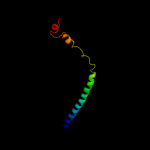
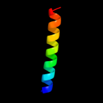
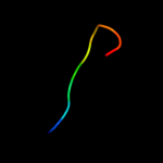
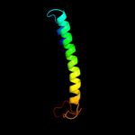
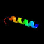
Image coloured by rainbow N → C terminus
Model (left) based on template d1f6ga_
Top template information
Fold:Voltage-gated potassium channels
Superfamily:Voltage-gated potassium channels
Family:Voltage-gated potassium channels
Confidence and coverage
Confidence: 51.8% Coverage: 58%
69 residues ( 58% of your sequence) have been modelled with 51.8% confidence by the single highest scoring template.
You may wish to submit your sequence to Phyrealarm. This will automatically scan your sequence every week for new potential templates as they appear in the Phyre2 library.
Please note: You must be registered and logged in to use Phyrealarm.
3D viewing
Interactive 3D view in Jmol

Sequence analysis 

Secondary structure and disorder prediction 

   1........10.........20.........30.........40.........50.........60
Sequence  MAGINLNKKLWRIPQQGMVRGVCAGIANYFDVPVKLVRILVVLSIFFGLALFTLVAYIIL
Secondary structure 


SS confidence 


Disorder  ????????


?


Disorder confidence 


   .........70.........80.........90.........100.........110.........
Sequence  SFALDPMPDNMAFGEQLPSSSELLDEVDRELAASETRLREMERYVTSDTFTLRSRFRQL
Secondary structure 


SS confidence 


Disorder 


????????????


?


???
Disorder confidence 


Confidence Key
High(9)                    Low (0)
?Disordered
Alpha helix
Beta strand

Domain analysis 

Hover over an aligned region to see model and summary info

Please note, only up to the top 20 hits are modelled to reduce computer load

RankAligned region

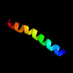
PDB 1f6g chain A

3D model

Region: 35 - 103
Aligned: 69
Modelled: 69
Confidence: 51.8%
Identity: 13%
Fold: Voltage-gated potassium channels
Superfamily: Voltage-gated potassium channels
Family: Voltage-gated potassium channels

Phyre2

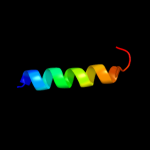
PDB 3r1f chain O

3D model

Region: 22 - 34
Aligned: 13
Modelled: 13
Confidence: 39.1%
Identity: 31%
PDB header:transcription
Chain: O: PDB Molecule:esx-1 secretion-associated regulator espr;
PDBTitle: crystal structure of a key regulator of virulence in mycobacterium2 tuberculosis

Phyre2

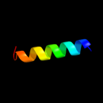
PDB 3okq chain A

3D model

Region: 75 - 114
Aligned: 40
Modelled: 40
Confidence: 23.3%
Identity: 8%
PDB header:protein binding
Chain: A: PDB Molecule:bud site selection protein 6;
PDBTitle: crystal structure of a core domain of yeast actin nucleation cofactor2 bud6

Phyre2

PDB 2o0c chain B

3D model

Region: 11 - 34
Aligned: 24
Modelled: 24
Confidence: 18.1%
Identity: 21%
PDB header:signaling protein
Chain: B: PDB Molecule:alr2278 protein;
PDBTitle: crystal structure of the h-nox domain from nostoc sp. pcc 71202 complexed to no

Phyre2

PDB 3pp5 chain A

3D model

Region: 83 - 107
Aligned: 25
Modelled: 25
Confidence: 14.7%
Identity: 12%
PDB header:structural protein
Chain: A: PDB Molecule:brk1;
PDBTitle: high-resolution structure of the trimeric scar/wave complex precursor2 brk1

Phyre2

PDB 1v54 chain J

3D model

Region: 38 - 67
Aligned: 30
Modelled: 30
Confidence: 13.9%
Identity: 13%
Fold: Single transmembrane helix
Superfamily: Mitochondrial cytochrome c oxidase subunit VIIa
Family: Mitochondrial cytochrome c oxidase subunit VIIa

Phyre2

PDB 1htm chain B

3D model

Region: 82 - 105
Aligned: 24
Modelled: 24
Confidence: 12.3%
Identity: 29%
PDB header:viral protein
Chain: B: PDB Molecule:hemagglutinin ha2 chain;
PDBTitle: structure of influenza haemagglutinin at the ph of membrane2 fusion

Phyre2

PDB 3e11 chain A domain 1

3D model

Region: 26 - 34
Aligned: 9
Modelled: 9
Confidence: 12.0%
Identity: 33%
Fold: Zincin-like
Superfamily: Metalloproteases ("zincins"), catalytic domain
Family: TTHA0227-like

Phyre2

PDB 1zgx chain B

3D model

Region: 101 - 110
Aligned: 10
Modelled: 10
Confidence: 10.8%
Identity: 40%
PDB header:hydrolase
Chain: B: PDB Molecule:guanyl-specific ribonuclease sa;
PDBTitle: crystal structure of ribonuclease mutant

Phyre2

PDB 1emz chain A

3D model

Region: 21 - 29
Aligned: 9
Modelled: 9
Confidence: 10.0%
Identity: 67%
PDB header:viral protein
Chain: A: PDB Molecule:envelope glycoprotein e1;
PDBTitle: solution structure of fragment (350-370) of the2 transmembrane domain of hepatitis c envelope glycoprotein3 e1

Phyre2

PDB 3f5d chain A

3D model

Region: 14 - 25
Aligned: 12
Modelled: 12
Confidence: 9.4%
Identity: 8%
PDB header:structural genomics, unknown function
Chain: A: PDB Molecule:protein ydea;
PDBTitle: crystal structure of a protein of unknown function from2 bacillus subtilis

Phyre2

PDB 2vvy chain C

3D model

Region: 21 - 48
Aligned: 28
Modelled: 28
Confidence: 9.3%
Identity: 29%
PDB header:viral protein
Chain: C: PDB Molecule:protein b15;
PDBTitle: structure of vaccinia virus protein b14

Phyre2

PDB 3hd6 chain A

3D model

Region: 17 - 78
Aligned: 62
Modelled: 62
Confidence: 8.8%
Identity: 13%
PDB header:membrane protein, transport protein
Chain: A: PDB Molecule:ammonium transporter rh type c;
PDBTitle: crystal structure of the human rhesus glycoprotein rhcg

Phyre2

PDB 1nhl chain A

3D model

Region: 81 - 107
Aligned: 27
Modelled: 27
Confidence: 8.5%
Identity: 26%
PDB header:protein transport
Chain: A: PDB Molecule:synaptosomal-associated protein 23;
PDBTitle: snap-23n structure

Phyre2

PDB 1txp chain A

3D model

Region: 84 - 103
Aligned: 20
Modelled: 20
Confidence: 8.1%
Identity: 15%
PDB header:signaling protein
Chain: A: PDB Molecule:heterogeneous nuclear ribonucleoprotein c
PDBTitle: heterogeneous nuclear ribonucleoprotein (hnrnp) c2 oligomerization domain tetramer

Phyre2

PDB 1txp chain B

3D model

Region: 84 - 103
Aligned: 20
Modelled: 20
Confidence: 8.1%
Identity: 15%
PDB header:signaling protein
Chain: B: PDB Molecule:heterogeneous nuclear ribonucleoprotein c
PDBTitle: heterogeneous nuclear ribonucleoprotein (hnrnp) c2 oligomerization domain tetramer

Phyre2

PDB 1txp chain D

3D model

Region: 84 - 103
Aligned: 20
Modelled: 20
Confidence: 8.1%
Identity: 15%
PDB header:signaling protein
Chain: D: PDB Molecule:heterogeneous nuclear ribonucleoprotein c
PDBTitle: heterogeneous nuclear ribonucleoprotein (hnrnp) c2 oligomerization domain tetramer

Phyre2

PDB 1txp chain C

3D model

Region: 84 - 103
Aligned: 20
Modelled: 20
Confidence: 8.1%
Identity: 15%
PDB header:signaling protein
Chain: C: PDB Molecule:heterogeneous nuclear ribonucleoprotein c
PDBTitle: heterogeneous nuclear ribonucleoprotein (hnrnp) c2 oligomerization domain tetramer

Phyre2

PDB 3swf chain A

3D model

Region: 82 - 119
Aligned: 38
Modelled: 38
Confidence: 7.6%
Identity: 16%
PDB header:transport protein
Chain: A: PDB Molecule:cgmp-gated cation channel alpha-1;
PDBTitle: cnga1 621-690 containing clz domain

Phyre2

PDB 2y0n chain D

3D model

Region: 22 - 31
Aligned: 10
Modelled: 10
Confidence: 7.3%
Identity: 30%
PDB header:transcription
Chain: D: PDB Molecule:male-specific lethal 3 homolog;
PDBTitle: crystal structure of the complex between dosage2 compensation factors msl1 and msl3

Phyre2
1

d1f6ga_
2

c3r1fO_
3

c3okqA_
4

c2o0cB_
5

c3pp5A_
6

d1v54j_
7

c1htmB_
8

d3e11a1
9

c1zgxB_
10

c1emzA_
11

c3f5dA_
12

c2vvyC_
13

c3hd6A_
14

c1nhlA_
15

c1txpA_
16

c1txpB_
17

c1txpD_
18

c1txpC_
19

c3swfA_
20

c2y0nD_
21


22


23


24


25


26


27


28


29


30


31


32


33


34


Detailed template information 

#
Template Alignment Coverage3D Model Confidence
% i.d. Template Information
1d1f6ga_


51.8 13 Fold:Voltage-gated potassium channels
Superfamily:Voltage-gated potassium channels
Family:Voltage-gated potassium channels
2c3r1fO_


39.1 31 PDB header:transcription
Chain: O: PDB Molecule:esx-1 secretion-associated regulator espr;
PDBTitle: crystal structure of a key regulator of virulence in mycobacterium2 tuberculosis
3c3okqA_


23.3 8 PDB header:protein binding
Chain: A: PDB Molecule:bud site selection protein 6;
PDBTitle: crystal structure of a core domain of yeast actin nucleation cofactor2 bud6
4c2o0cB_


18.1 21 PDB header:signaling protein
Chain: B: PDB Molecule:alr2278 protein;
PDBTitle: crystal structure of the h-nox domain from nostoc sp. pcc 71202 complexed to no
5c3pp5A_


14.7 12 PDB header:structural protein
Chain: A: PDB Molecule:brk1;
PDBTitle: high-resolution structure of the trimeric scar/wave complex precursor2 brk1
6d1v54j_


13.9 13 Fold:Single transmembrane helix
Superfamily:Mitochondrial cytochrome c oxidase subunit VIIa
Family:Mitochondrial cytochrome c oxidase subunit VIIa
7c1htmB_


12.3 29 PDB header:viral protein
Chain: B: PDB Molecule:hemagglutinin ha2 chain;
PDBTitle: structure of influenza haemagglutinin at the ph of membrane2 fusion
8d3e11a1


12.0 33 Fold:Zincin-like
Superfamily:Metalloproteases ("zincins"), catalytic domain
Family:TTHA0227-like
9c1zgxB_


10.8 40 PDB header:hydrolase
Chain: B: PDB Molecule:guanyl-specific ribonuclease sa;
PDBTitle: crystal structure of ribonuclease mutant
10c1emzA_


10.0 67 PDB header:viral protein
Chain: A: PDB Molecule:envelope glycoprotein e1;
PDBTitle: solution structure of fragment (350-370) of the2 transmembrane domain of hepatitis c envelope glycoprotein3 e1
11c3f5dA_


9.4 8 PDB header:structural genomics, unknown function
Chain: A: PDB Molecule:protein ydea;
PDBTitle: crystal structure of a protein of unknown function from2 bacillus subtilis
12c2vvyC_


9.3 29 PDB header:viral protein
Chain: C: PDB Molecule:protein b15;
PDBTitle: structure of vaccinia virus protein b14
13c3hd6A_


8.8 13 PDB header:membrane protein, transport protein
Chain: A: PDB Molecule:ammonium transporter rh type c;
PDBTitle: crystal structure of the human rhesus glycoprotein rhcg
14c1nhlA_


8.5 26 PDB header:protein transport
Chain: A: PDB Molecule:synaptosomal-associated protein 23;
PDBTitle: snap-23n structure
15c1txpA_


8.1 15 PDB header:signaling protein
Chain: A: PDB Molecule:heterogeneous nuclear ribonucleoprotein c
PDBTitle: heterogeneous nuclear ribonucleoprotein (hnrnp) c2 oligomerization domain tetramer
16c1txpB_


8.1 15 PDB header:signaling protein
Chain: B: PDB Molecule:heterogeneous nuclear ribonucleoprotein c
PDBTitle: heterogeneous nuclear ribonucleoprotein (hnrnp) c2 oligomerization domain tetramer
17c1txpD_


8.1 15 PDB header:signaling protein
Chain: D: PDB Molecule:heterogeneous nuclear ribonucleoprotein c
PDBTitle: heterogeneous nuclear ribonucleoprotein (hnrnp) c2 oligomerization domain tetramer
18c1txpC_


8.1 15 PDB header:signaling protein
Chain: C: PDB Molecule:heterogeneous nuclear ribonucleoprotein c
PDBTitle: heterogeneous nuclear ribonucleoprotein (hnrnp) c2 oligomerization domain tetramer
19c3swfA_


7.6 16 PDB header:transport protein
Chain: A: PDB Molecule:cgmp-gated cation channel alpha-1;
PDBTitle: cnga1 621-690 containing clz domain
20c2y0nD_


7.3 30 PDB header:transcription
Chain: D: PDB Molecule:male-specific lethal 3 homolog;
PDBTitle: crystal structure of the complex between dosage2 compensation factors msl1 and msl3
21d1ukfa_


not modelled 7.1 27 Fold:Cysteine proteinases
Superfamily:Cysteine proteinases
Family:Avirulence protein Avrpph3
22c3rmsA_


not modelled 7.1 21 PDB header:structural genomics, unknown function
Chain: A: PDB Molecule:uncharacterized protein;
PDBTitle: crystal structure of uncharacterized protein svir_20580 from2 saccharomonospora viridis
23c2vvxA_


not modelled 7.1 19 PDB header:viral protein
Chain: A: PDB Molecule:protein a52;
PDBTitle: structure of vaccinia virus protein a52
24c3p8cE_


not modelled 7.0 30 PDB header:protein binding
Chain: E: PDB Molecule:probable protein brick1;
PDBTitle: structure and control of the actin regulatory wave complex
25d1r69a_


not modelled 6.9 25 Fold:lambda repressor-like DNA-binding domains
Superfamily:lambda repressor-like DNA-binding domains
Family:Phage repressors
26d2fexa1


not modelled 6.7 29 Fold:Flavodoxin-like
Superfamily:Class I glutamine amidotransferase-like
Family:DJ-1/PfpI
27d1pv0a_


not modelled 6.7 25 Fold:Long alpha-hairpin
Superfamily:Sporulation inhibitor Sda
Family:Sporulation inhibitor Sda
28c1envA_


not modelled 6.5 14 PDB header:viral protein
Chain: A: PDB Molecule:hiv-1 envelope protein chimera consisting of a fragment of
PDBTitle: atomic structure of the ectodomain from hiv-1 gp41
29c2k6sB_


not modelled 6.2 30 PDB header:protein transport
Chain: B: PDB Molecule:rab11fip2 protein;
PDBTitle: structure of rab11-fip2 c-terminal coiled-coil domain
30c3i5dC_


not modelled 6.2 19 PDB header:transport protein
Chain: C: PDB Molecule:p2x purinoceptor;
PDBTitle: crystal structure of the atp-gated p2x4 ion channel in the closed, apo2 state at 3.5 angstroms (r3)
31c2f5jA_


not modelled 6.1 60 PDB header:gene regulation
Chain: A: PDB Molecule:mortality factor 4-like protein 1;
PDBTitle: crystal structure of mrg domain from human mrg15
32c3cneD_


not modelled 5.8 36 PDB header:hydrolase
Chain: D: PDB Molecule:putative protease i;
PDBTitle: crystal structure of the putative protease i from bacteroides2 thetaiotaomicron
33c3efeC_


not modelled 5.8 18 PDB header:chaperone
Chain: C: PDB Molecule:thij/pfpi family protein;
PDBTitle: the crystal structure of the thij/pfpi family protein from bacillus2 anthracis
34d2axte1


not modelled 5.5 19 Fold:Single transmembrane helix
Superfamily:Cytochrome b559 subunits
Family:Cytochrome b559 subunits

Binding site prediction 

Due to computational demand, binding site predictions are not run for batch jobs

If you want to predict binding sites, please manually submit your model of choice to 3DLigandSite


Phyre is for academic use only

Please cite: Protein structure prediction on the web: a case study using the Phyre server
Kelley LA and Sternberg MJE. Nature Protocols 4, 363 - 371 (2009) [pdf] [Import into BibTeX]
 
If you use the binding site predictions from 3DLigandSite, please also cite:
3DLigandSite: predicting ligand-binding sites using similar structures.
Wass MN, Kelley LA and Sternberg MJ Nucleic Acids Research 38, W469-73 (2010) [PubMed]
 
© Structural Bioinformatics Group
Imperial College London
Lawrence Kelley, Benjamin Jefferys
Disclaimer
Terms and Conditions
Component software
Template detection: HHpred 1.51
Secondary structure prediction: Psi-pred 2.5
Disorder prediction: Disopred 2.4
Transmembrane prediction: Memsat_SVM
Multi-template modelling and ab initio: Poing 1.0