| Secondary structure and disorder prediction | |
| | |
1 | . | . | . | . | . | . | . | . | 10 | . | . | . | . | . | . | . | . | . | 20 | . | . | . | . | . | . | . | . | . | 30 | . | . | . | . | . | . | . | . | . | 40 | . | . | . | . | . | . | . | . | . | 50 | . | . | . | . | . | . | . | . | . | 60 |
| Sequence | |
M | A | Q | F | E | W | V | H | A | A | W | L | A | L | A | I | V | L | E | I | V | A | N | V | F | L | K | F | S | D | G | F | R | R | K | I | F | G | L | L | S | L | A | A | V | L | A | A | F | S | A | L | S | Q | A | V | K | G | I | D |
| Secondary structure | |
|
|
|  | | | | | | | | | | | | | | | | | | | | | | | | | |
|
|
|
|
|
| | | | | | | | | | | | | | | | | | | | | | | | |
|
| SS confidence | |
|
|
|
|
|
|
|
|
|
|
|
|
|
|
|
|
|
|
|
|
|
|
|
|
|
|
|
|
|
|
|
|
|
|
|
|
|
|
|
|
|
|
|
|
|
|
|
|
|
|
|
|
|
|
|
|
|
|
|
|
| Disorder | |
? | ? | ? | ? | ? | ? |
|
|
|
|
|
|
|
|
|
|
|
|
|
|
|
|
|
|
|
|
|
|
| ? | ? |
|
|
|
|
|
|
|
|
|
|
|
|
|
|
|
|
|
|
|
|
|
|
|
|
| ? |
|
|
|
| Disorder confidence | |
|
|
|
|
|
|
|
|
|
|
|
|
|
|
|
|
|
|
|
|
|
|
|
|
|
|
|
|
|
|
|
|
|
|
|
|
|
|
|
|
|
|
|
|
|
|
|
|
|
|
|
|
|
|
|
|
|
|
|
|
| |
| | |
. | . | . | . | . | . | . | . | . | 70 | . | . | . | . | . | . | . | . | . | 80 | . | . | . | . | . | . | . | . | . | 90 | . | . | . | . | . | . | . | . | . | 100 | . | . | . | . | . | . | . | . | . |
| Sequence | |
L | S | V | A | Y | A | L | W | G | G | F | G | I | A | A | T | L | A | A | G | W | I | L | F | G | Q | R | L | N | R | K | G | W | I | G | L | V | L | L | L | A | G | M | I | M | V | K | L | A |
| Secondary structure | |
| | | | | | | | | | | | | | | | | | | | | | | |
|
|
|
|
| | | | | | | | | | | | | | | | | | |
|
|
| SS confidence | |
|
|
|
|
|
|
|
|
|
|
|
|
|
|
|
|
|
|
|
|
|
|
|
|
|
|
|
|
|
|
|
|
|
|
|
|
|
|
|
|
|
|
|
|
|
|
|
|
|
| Disorder | |
|
|
|
|
|
|
|
|
|
|
|
|
|
|
|
|
|
|
|
|
|
|
|
|
|
|
|
|
|
|
|
|
|
|
|
|
|
|
|
|
|
|
|
|
|
| ? | ? | ? |
| Disorder confidence | |
|
|
|
|
|
|
|
|
|
|
|
|
|
|
|
|
|
|
|
|
|
|
|
|
|
|
|
|
|
|
|
|
|
|
|
|
|
|
|
|
|
|
|
|
|
|
|
|
|
| |
| Confidence Key |
| High(9) | |
|
|
|
|
|
|
|
|
|
Low (0) |
| ? | Disordered |
| Alpha helix |
  | Beta strand |
Hover over an aligned region to see model and summary info
Please note, only up to the top 20 hits are modelled to reduce computer load
|
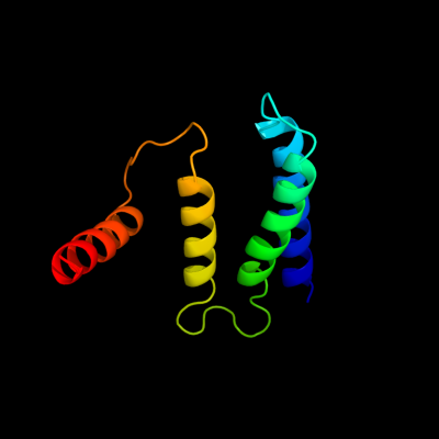
| 1 |
|
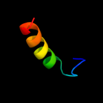
PDB 1s7b chain A
Region: 11 - 108
Aligned: 98
Modelled: 98
Confidence: 100.0%
Identity: 35%
Fold: Multidrug resistance efflux transporter EmrE
Superfamily: Multidrug resistance efflux transporter EmrE
Family: Multidrug resistance efflux transporter EmrE
Phyre2
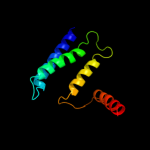
| 2 |
|
PDB 2i68 chain B
Region: 9 - 108
Aligned: 77
Modelled: 88
Confidence: 99.8%
Identity: 31%
PDB header:transport protein
Chain: B: PDB Molecule:protein emre;
PDBTitle: cryo-em based theoretical model structure of transmembrane2 domain of the multidrug-resistance antiporter from e. coli3 emre
Phyre2
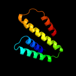
| 3 |
|
PDB 3mp7 chain B
Region: 90 - 106
Aligned: 17
Modelled: 17
Confidence: 14.7%
Identity: 29%
PDB header:protein transport
Chain: B: PDB Molecule:preprotein translocase subunit sece;
PDBTitle: lateral opening of a translocon upon entry of protein suggests the2 mechanism of insertion into membranes
Phyre2
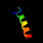
| 4 |
|
PDB 2vqc chain A
Region: 54 - 75
Aligned: 22
Modelled: 22
Confidence: 7.6%
Identity: 18%
PDB header:dna-binding protein
Chain: A: PDB Molecule:hypothetical 13.2 kda protein;
PDBTitle: structure of a dna binding winged-helix protein, f-112,2 from sulfolobus spindle-shaped virus 1.
Phyre2
| 5 |
|
PDB 2vqc chain A domain 1
Region: 54 - 75
Aligned: 22
Modelled: 22
Confidence: 7.6%
Identity: 18%
Fold: DNA/RNA-binding 3-helical bundle
Superfamily: "Winged helix" DNA-binding domain
Family: F112-like
Phyre2
| 6 |
|
PDB 2axt chain J domain 1
Region: 72 - 85
Aligned: 14
Modelled: 14
Confidence: 6.2%
Identity: 21%
Fold: Single transmembrane helix
Superfamily: Photosystem II reaction center protein J, PsbJ
Family: PsbJ-like
Phyre2
|
| Detailed template information | |
Due to computational demand, binding site predictions are not run for batch jobs
If you want to predict binding sites, please manually submit your model of choice to 3DLigandSite
Phyre is for academic use only
| Please cite: Protein structure prediction on
the web: a case study using the Phyre server |
| Kelley LA and Sternberg MJE. Nature Protocols
4, 363 - 371 (2009) [pdf] [Import into BibTeX] |
| |
| If you use the binding site
predictions from 3DLigandSite, please also cite: |
| 3DLigandSite: predicting ligand-binding sites using similar structures. |
| Wass MN, Kelley LA and Sternberg
MJ Nucleic Acids Research 38, W469-73 (2010) [PubMed] |
| |
|
|
|
|