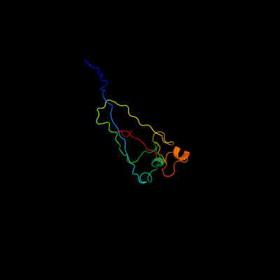
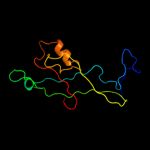
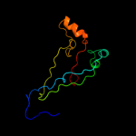
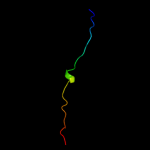
| 1 |
|
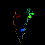
PDB 3jwn chain K
Region: 29 - 141
Aligned: 98
Modelled: 113
Confidence: 97.4%
Identity: 17%
PDB header:protein binding/cell adhesion
Chain: K: PDB Molecule:protein fimf;
PDBTitle: complex of fimc, fimf, fimg and fimh
Phyre2
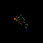
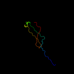
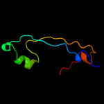
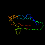
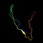
| 2 |
|
PDB 3jwn chain L
Region: 29 - 141
Aligned: 98
Modelled: 113
Confidence: 97.3%
Identity: 17%
PDB header:protein binding/cell adhesion
Chain: L: PDB Molecule:protein fimf;
PDBTitle: complex of fimc, fimf, fimg and fimh
Phyre2
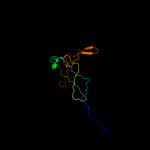
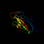
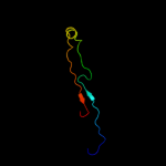
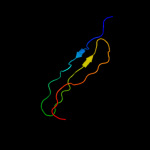
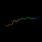
| 3 |
|
PDB 3jwn chain E
Region: 29 - 141
Aligned: 98
Modelled: 113
Confidence: 97.3%
Identity: 17%
PDB header:protein binding/cell adhesion
Chain: E: PDB Molecule:protein fimf;
PDBTitle: complex of fimc, fimf, fimg and fimh
Phyre2
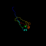
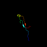
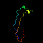
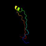
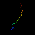
| 4 |
|
PDB 2jty chain A
Region: 24 - 141
Aligned: 105
Modelled: 118
Confidence: 97.2%
Identity: 14%
PDB header:structural protein
Chain: A: PDB Molecule:type-1 fimbrial protein, a chain;
PDBTitle: self-complemented variant of fima, the main subunit of type 1 pilus
Phyre2
| 5 |
|
PDB 3jwn chain F
Region: 29 - 104
Aligned: 71
Modelled: 76
Confidence: 97.1%
Identity: 18%
PDB header:protein binding/cell adhesion
Chain: F: PDB Molecule:protein fimf;
PDBTitle: complex of fimc, fimf, fimg and fimh
Phyre2
| 6 |
|
PDB 2w07 chain B
Region: 39 - 171
Aligned: 119
Modelled: 125
Confidence: 96.1%
Identity: 15%
PDB header:cell adhesion
Chain: B: PDB Molecule:minor pilin subunit papf;
PDBTitle: structural determinants of polymerization reactivity of the2 p pilus adaptor subunit papf
Phyre2
| 7 |
|
PDB 2uy6 chain B domain 1
Region: 32 - 91
Aligned: 54
Modelled: 54
Confidence: 95.0%
Identity: 17%
Fold: Common fold of diphtheria toxin/transcription factors/cytochrome f
Superfamily: Bacterial adhesins
Family: Pilus subunits
Phyre2
| 8 |
|
PDB 2jmr chain A
Region: 29 - 141
Aligned: 98
Modelled: 113
Confidence: 94.5%
Identity: 15%
PDB header:cell adhesion
Chain: A: PDB Molecule:fimf;
PDBTitle: nmr structure of the e. coli type 1 pilus subunit fimf
Phyre2
| 9 |
|
PDB 1n12 chain A
Region: 42 - 99
Aligned: 48
Modelled: 58
Confidence: 94.0%
Identity: 29%
Fold: Common fold of diphtheria toxin/transcription factors/cytochrome f
Superfamily: Bacterial adhesins
Family: Pilus subunits
Phyre2
| 10 |
|
PDB 2j2z chain B domain 1
Region: 33 - 90
Aligned: 52
Modelled: 52
Confidence: 94.0%
Identity: 19%
Fold: Common fold of diphtheria toxin/transcription factors/cytochrome f
Superfamily: Bacterial adhesins
Family: Pilus subunits
Phyre2
| 11 |
|
PDB 3bfw chain A
Region: 42 - 98
Aligned: 51
Modelled: 57
Confidence: 92.9%
Identity: 24%
PDB header:structural protein/structural protein
Chain: A: PDB Molecule:protein fimg;
PDBTitle: crystal structure of truncated fimg (fimgt) in complex with the donor2 strand peptide of fimf (dsf)
Phyre2
| 12 |
|
PDB 2wmp chain B
Region: 42 - 134
Aligned: 64
Modelled: 75
Confidence: 92.5%
Identity: 27%
PDB header:chaperone
Chain: B: PDB Molecule:papg protein;
PDBTitle: structure of the e. coli chaperone papd in complex with the pilin2 domain of the papgii adhesin
Phyre2
| 13 |
|
PDB 1ze3 chain H domain 1
Region: 42 - 142
Aligned: 74
Modelled: 85
Confidence: 91.7%
Identity: 16%
Fold: Common fold of diphtheria toxin/transcription factors/cytochrome f
Superfamily: Bacterial adhesins
Family: Pilus subunits
Phyre2
| 14 |
|
PDB 1klf chain P
Region: 38 - 104
Aligned: 50
Modelled: 51
Confidence: 89.7%
Identity: 14%
PDB header:chaperone/adhesin complex
Chain: P: PDB Molecule:fimh protein;
PDBTitle: fimh adhesin-fimc chaperone complex with d-mannose
Phyre2
| 15 |
|
PDB 1pdk chain B
Region: 39 - 90
Aligned: 47
Modelled: 52
Confidence: 85.7%
Identity: 4%
Fold: Common fold of diphtheria toxin/transcription factors/cytochrome f
Superfamily: Bacterial adhesins
Family: Pilus subunits
Phyre2
| 16 |
|
PDB 2jna chain A domain 1
Region: 3 - 30
Aligned: 26
Modelled: 28
Confidence: 13.3%
Identity: 35%
Fold: Dodecin subunit-like
Superfamily: YdgH-like
Family: YdgH-like
Phyre2
| 17 |
|
PDB 3qbt chain H
Region: 45 - 104
Aligned: 45
Modelled: 46
Confidence: 8.6%
Identity: 9%
PDB header:protein transport/hydrolase
Chain: H: PDB Molecule:inositol polyphosphate 5-phosphatase ocrl-1;
PDBTitle: crystal structure of ocrl1 540-678 in complex with rab8a:gppnhp
Phyre2
| 18 |
|
PDB 2kr7 chain A
Region: 212 - 234
Aligned: 23
Modelled: 23
Confidence: 8.2%
Identity: 13%
PDB header:isomerase
Chain: A: PDB Molecule:fkbp-type peptidyl-prolyl cis-trans isomerase slyd;
PDBTitle: solution structure of helicobacter pylori slyd
Phyre2
| 19 |
|
PDB 2kfw chain A
Region: 211 - 225
Aligned: 15
Modelled: 15
Confidence: 7.2%
Identity: 13%
PDB header:isomerase
Chain: A: PDB Molecule:fkbp-type peptidyl-prolyl cis-trans isomerase
PDBTitle: solution structure of full-length slyd from e.coli
Phyre2
| 20 |
|
PDB 3klq chain B
Region: 41 - 60
Aligned: 20
Modelled: 20
Confidence: 7.0%
Identity: 10%
PDB header:cell adhesion
Chain: B: PDB Molecule:putative pilus anchoring protein;
PDBTitle: crystal structure of the minor pilin fctb from streptococcus pyogenes2 90/306s
Phyre2
| 21 |
|