Phyre Home Retrieve Phyre Job Id
Emaill.a.kelley@imperial.ac.uk
DescriptionQ47268
DateThu Jan 5 12:36:40 GMT 2012
Unique Job IDce46cbb2c157167d

Summary 

Top model
Image coloured by rainbow N → C terminus
Model (left) based on template d1e4va2
Top template information
Fold:Rubredoxin-like
Superfamily:Microbial and mitochondrial ADK, insert "zinc finger" domain
Family:Microbial and mitochondrial ADK, insert "zinc finger" domain
Confidence and coverage
Confidence: 7.5% Coverage: 67%
22 residues ( 67% of your sequence) have been modelled with 7.5% confidence by the single highest scoring template.
You may wish to submit your sequence to Phyrealarm. This will automatically scan your sequence every week for new potential templates as they appear in the Phyre2 library.
Please note: You must be registered and logged in to use Phyrealarm.
3D viewing
Interactive 3D view in Jmol

Sequence analysis 

Secondary structure and disorder prediction 

   1........10.........20.........30...
Sequence  MRTYNPNSLLPSQMQKCTCNSLHLAFDLCGGEA
Secondary structure 


SS confidence 


Disorder  ???????


??


??????
Disorder confidence 


Confidence Key
High(9)                    Low (0)
?Disordered
Alpha helix
Beta strand

Domain analysis 

Hover over an aligned region to see model and summary info

Please note, only up to the top 20 hits are modelled to reduce computer load

RankAligned region

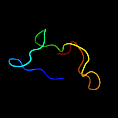
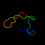
PDB 1e4v chain A domain 2

3D model

Region: 2 - 32
Aligned: 22
Modelled: 31
Confidence: 7.5%
Identity: 32%
Fold: Rubredoxin-like
Superfamily: Microbial and mitochondrial ADK, insert "zinc finger" domain
Family: Microbial and mitochondrial ADK, insert "zinc finger" domain

Phyre2

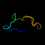
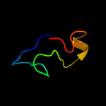
PDB 1p3j chain A domain 2

3D model

Region: 2 - 32
Aligned: 22
Modelled: 31
Confidence: 7.1%
Identity: 41%
Fold: Rubredoxin-like
Superfamily: Microbial and mitochondrial ADK, insert "zinc finger" domain
Family: Microbial and mitochondrial ADK, insert "zinc finger" domain

Phyre2

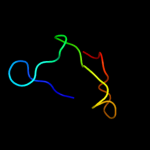
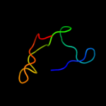
PDB 2ak3 chain A domain 2

3D model

Region: 2 - 32
Aligned: 22
Modelled: 31
Confidence: 7.0%
Identity: 36%
Fold: Rubredoxin-like
Superfamily: Microbial and mitochondrial ADK, insert "zinc finger" domain
Family: Microbial and mitochondrial ADK, insert "zinc finger" domain

Phyre2

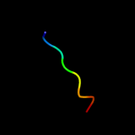
PDB 1zin chain A domain 2

3D model

Region: 2 - 32
Aligned: 22
Modelled: 31
Confidence: 6.4%
Identity: 45%
Fold: Rubredoxin-like
Superfamily: Microbial and mitochondrial ADK, insert "zinc finger" domain
Family: Microbial and mitochondrial ADK, insert "zinc finger" domain

Phyre2

PDB 1s3g chain A domain 2

3D model

Region: 2 - 32
Aligned: 22
Modelled: 31
Confidence: 6.4%
Identity: 41%
Fold: Rubredoxin-like
Superfamily: Microbial and mitochondrial ADK, insert "zinc finger" domain
Family: Microbial and mitochondrial ADK, insert "zinc finger" domain

Phyre2

PDB 1vs6 chain 2

3D model

Region: 2 - 9
Aligned: 8
Modelled: 8
Confidence: 5.7%
Identity: 50%
PDB header:ribosome
Chain: 2: PDB Molecule:50s ribosomal protein l34;
PDBTitle: crystal structure of the bacterial ribosome from2 escherichia coli in complex with the antibiotic kasugamyin3 at 3.5a resolution. this file contains the 50s subunit of4 one 70s ribosome. the entire crystal structure contains5 two 70s ribosomes and is described in remark 400.

Phyre2

PDB 3e1d chain V

3D model

Region: 2 - 9
Aligned: 8
Modelled: 8
Confidence: 5.7%
Identity: 50%
PDB header:ribosome
Chain: V: PDB Molecule:50s ribosomal protein l34;
PDBTitle: structure of the 50s subunit of e. coli ribosome in post-2 accommodation state

Phyre2

PDB 1vs8 chain 2

3D model

Region: 2 - 9
Aligned: 8
Modelled: 8
Confidence: 5.6%
Identity: 50%
PDB header:ribosome
Chain: 2: PDB Molecule:50s ribosomal protein l34;
PDBTitle: crystal structure of the bacterial ribosome from escherichia coli in2 complex with the antibiotic kasugamyin at 3.5a resolution. this file3 contains the 50s subunit of one 70s ribosome. the entire crystal4 structure contains two 70s ribosomes and is described in remark 400.

Phyre2

PDB 2awb chain 2

3D model

Region: 2 - 9
Aligned: 8
Modelled: 8
Confidence: 5.6%
Identity: 50%
PDB header:ribosome
Chain: 2: PDB Molecule:50s ribosomal protein l34;
PDBTitle: crystal structure of the bacterial ribosome from2 escherichia coli at 3.5 a resolution. this file contains3 the 50s subunit of the second 70s ribosome. the entire4 crystal structure contains two 70s ribosomes and is5 described in remark 400.

Phyre2

PDB 2j28 chain 2

3D model

Region: 2 - 9
Aligned: 8
Modelled: 8
Confidence: 5.6%
Identity: 50%
PDB header:ribosome
Chain: 2: PDB Molecule:50s ribosomal protein l34;
PDBTitle: model of e. coli srp bound to 70s rncs

Phyre2

PDB 3e1b chain V

3D model

Region: 2 - 9
Aligned: 8
Modelled: 8
Confidence: 5.6%
Identity: 50%
PDB header:ribosome
Chain: V: PDB Molecule:50s ribosomal protein l34;
PDBTitle: structure of the 50s subunit of e. coli ribosome in pre-2 accommodation state

Phyre2

PDB 2rdo chain 2

3D model

Region: 2 - 9
Aligned: 8
Modelled: 8
Confidence: 5.6%
Identity: 50%
PDB header:ribosome
Chain: 2: PDB Molecule:50s ribosomal protein l34;
PDBTitle: 50s subunit with ef-g(gdpnp) and rrf bound

Phyre2

PDB 2aw4 chain 2

3D model

Region: 2 - 9
Aligned: 8
Modelled: 8
Confidence: 5.6%
Identity: 50%
PDB header:ribosome
Chain: 2: PDB Molecule:50s ribosomal protein l34;
PDBTitle: crystal structure of the bacterial ribosome from2 escherichia coli at 3.5 a resolution. this file contains3 the 50s subunit of one 70s ribosome. the entire crystal4 structure contains two 70s ribosomes and is described in5 remark 400.

Phyre2

PDB 3bbx chain 2

3D model

Region: 2 - 9
Aligned: 8
Modelled: 8
Confidence: 5.6%
Identity: 50%
PDB header:ribosome
Chain: 2: PDB Molecule:50s ribosomal protein l34;
PDBTitle: the hsp15 protein fitted into the low resolution cryo-em map of the2 50s.nc-trna.hsp15 complex

Phyre2

PDB 3izt chain E

3D model

Region: 2 - 9
Aligned: 8
Modelled: 8
Confidence: 5.2%
Identity: 50%
PDB header:ribosome
Chain: E: PDB Molecule:50s ribosomal protein l3;
PDBTitle: structural insights into cognate vs. near-cognate discrimination2 during decoding. this entry contains the large subunit of a ribosome3 programmed with a near-cognate codon.

Phyre2
1

d1e4va2
2

d1p3ja2
3

d2ak3a2
4

d1zina2
5

d1s3ga2
6

c1vs62_
7

c3e1dV_
8

c1vs82_
9

c2awb2_
10

c2j282_
11

c3e1bV_
12

c2rdo2_
13

c2aw42_
14

c3bbx2_
15

c3izte_


Detailed template information 

#
Template Alignment Coverage3D Model Confidence
% i.d. Template Information
1d1e4va2


7.5 32 Fold:Rubredoxin-like
Superfamily:Microbial and mitochondrial ADK, insert "zinc finger" domain
Family:Microbial and mitochondrial ADK, insert "zinc finger" domain
2d1p3ja2


7.1 41 Fold:Rubredoxin-like
Superfamily:Microbial and mitochondrial ADK, insert "zinc finger" domain
Family:Microbial and mitochondrial ADK, insert "zinc finger" domain
3d2ak3a2


7.0 36 Fold:Rubredoxin-like
Superfamily:Microbial and mitochondrial ADK, insert "zinc finger" domain
Family:Microbial and mitochondrial ADK, insert "zinc finger" domain
4d1zina2


6.4 45 Fold:Rubredoxin-like
Superfamily:Microbial and mitochondrial ADK, insert "zinc finger" domain
Family:Microbial and mitochondrial ADK, insert "zinc finger" domain
5d1s3ga2


6.4 41 Fold:Rubredoxin-like
Superfamily:Microbial and mitochondrial ADK, insert "zinc finger" domain
Family:Microbial and mitochondrial ADK, insert "zinc finger" domain
6c1vs62_


5.7 50 PDB header:ribosome
Chain: 2: PDB Molecule:50s ribosomal protein l34;
PDBTitle: crystal structure of the bacterial ribosome from2 escherichia coli in complex with the antibiotic kasugamyin3 at 3.5a resolution. this file contains the 50s subunit of4 one 70s ribosome. the entire crystal structure contains5 two 70s ribosomes and is described in remark 400.
7c3e1dV_


5.7 50 PDB header:ribosome
Chain: V: PDB Molecule:50s ribosomal protein l34;
PDBTitle: structure of the 50s subunit of e. coli ribosome in post-2 accommodation state
8c1vs82_


5.6 50 PDB header:ribosome
Chain: 2: PDB Molecule:50s ribosomal protein l34;
PDBTitle: crystal structure of the bacterial ribosome from escherichia coli in2 complex with the antibiotic kasugamyin at 3.5a resolution. this file3 contains the 50s subunit of one 70s ribosome. the entire crystal4 structure contains two 70s ribosomes and is described in remark 400.
9c2awb2_


5.6 50 PDB header:ribosome
Chain: 2: PDB Molecule:50s ribosomal protein l34;
PDBTitle: crystal structure of the bacterial ribosome from2 escherichia coli at 3.5 a resolution. this file contains3 the 50s subunit of the second 70s ribosome. the entire4 crystal structure contains two 70s ribosomes and is5 described in remark 400.
10c2j282_


5.6 50 PDB header:ribosome
Chain: 2: PDB Molecule:50s ribosomal protein l34;
PDBTitle: model of e. coli srp bound to 70s rncs
11c3e1bV_


5.6 50 PDB header:ribosome
Chain: V: PDB Molecule:50s ribosomal protein l34;
PDBTitle: structure of the 50s subunit of e. coli ribosome in pre-2 accommodation state
12c2rdo2_


5.6 50 PDB header:ribosome
Chain: 2: PDB Molecule:50s ribosomal protein l34;
PDBTitle: 50s subunit with ef-g(gdpnp) and rrf bound
13c2aw42_


5.6 50 PDB header:ribosome
Chain: 2: PDB Molecule:50s ribosomal protein l34;
PDBTitle: crystal structure of the bacterial ribosome from2 escherichia coli at 3.5 a resolution. this file contains3 the 50s subunit of one 70s ribosome. the entire crystal4 structure contains two 70s ribosomes and is described in5 remark 400.
14c3bbx2_


5.6 50 PDB header:ribosome
Chain: 2: PDB Molecule:50s ribosomal protein l34;
PDBTitle: the hsp15 protein fitted into the low resolution cryo-em map of the2 50s.nc-trna.hsp15 complex
15c3izte_


5.2 50 PDB header:ribosome
Chain: E: PDB Molecule:50s ribosomal protein l3;
PDBTitle: structural insights into cognate vs. near-cognate discrimination2 during decoding. this entry contains the large subunit of a ribosome3 programmed with a near-cognate codon.

Binding site prediction 

Due to computational demand, binding site predictions are not run for batch jobs

If you want to predict binding sites, please manually submit your model of choice to 3DLigandSite


Phyre is for academic use only

Please cite: Protein structure prediction on the web: a case study using the Phyre server
Kelley LA and Sternberg MJE. Nature Protocols 4, 363 - 371 (2009) [pdf] [Import into BibTeX]
 
If you use the binding site predictions from 3DLigandSite, please also cite:
3DLigandSite: predicting ligand-binding sites using similar structures.
Wass MN, Kelley LA and Sternberg MJ Nucleic Acids Research 38, W469-73 (2010) [PubMed]
 
© Structural Bioinformatics Group
Imperial College London
Lawrence Kelley, Benjamin Jefferys
Disclaimer
Terms and Conditions
Component software
Template detection: HHpred 1.51
Secondary structure prediction: Psi-pred 2.5
Disorder prediction: Disopred 2.4
Transmembrane prediction: Memsat_SVM
Multi-template modelling and ab initio: Poing 1.0