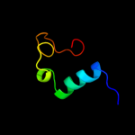
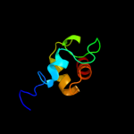
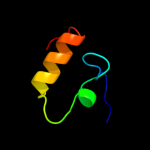
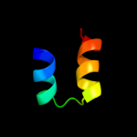
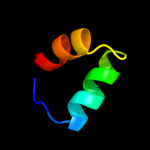
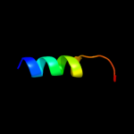
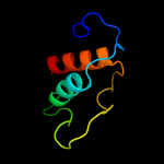
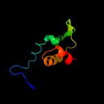
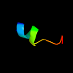
1 c2jvwA_
66.9
29
PDB header: structural genomics, unknown functionChain: A: PDB Molecule: uncharacterized protein;PDBTitle: solution nmr structure of uncharacterized protein q5e7h1 from vibrio2 fischeri. northeast structural genomics target vfr117
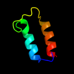
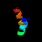
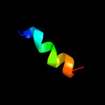
2 c2fwtA_
47.4
24
PDB header: electron transportChain: A: PDB Molecule: dhc, diheme cytochrome c;PDBTitle: crystal structure of dhc purified from rhodobacter2 sphaeroides
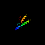
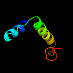
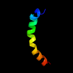
3 d1zeea1
40.5
33
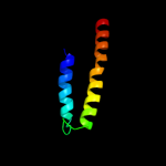
Fold: Indolic compounds 2,3-dioxygenase-likeSuperfamily: Indolic compounds 2,3-dioxygenase-likeFamily: Indoleamine 2,3-dioxygenase-like4 c1vngA_
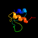
22.1
13
PDB header: haloperoxidaseChain: A: PDB Molecule: vanadium chloroperoxidase;PDBTitle: chloroperoxidase from the fungus curvularia inaequalis:2 mutant h404a
5 c1wr1B_
20.4
29
PDB header: signaling proteinChain: B: PDB Molecule: ubiquitin-like protein dsk2;PDBTitle: the complex sturcture of dsk2p uba with ubiquitin
6 c2hl7A_
19.9
24
PDB header: oxidoreductaseChain: A: PDB Molecule: cytochrome c-type biogenesis protein ccmh;PDBTitle: crystal structure of the periplasmic domain of ccmh from pseudomonas2 aeruginosa
7 c1yx3A_
18.4
18
PDB header: structural genomics, unknown functionChain: A: PDB Molecule: hypothetical protein dsrc;PDBTitle: nmr structure of allochromatium vinosum dsrc: northeast2 structural genomics consortium target op4
8 c2dnwA_
18.1
16
PDB header: transport proteinChain: A: PDB Molecule: acyl carrier protein;PDBTitle: solution structure of rsgi ruh-059, an acp domain of acyl2 carrier protein, mitochondrial [precursor] from human cdna
9 c3g2bA_
17.1
23
PDB header: biosynthetic proteinChain: A: PDB Molecule: coenzyme pqq synthesis protein d;PDBTitle: crystal structure of pqqd from xanthomonas campestris
10 c2kw0A_
16.6
20
PDB header: oxidoreductaseChain: A: PDB Molecule: ccmh protein;PDBTitle: solution structure of n-terminal domain of ccmh from escherichia.coli
11 d1wj7a1
16.1
18
Fold: RuvA C-terminal domain-likeSuperfamily: UBA-likeFamily: UBA domain12 c3hieA_
15.9
44
PDB header: exocytosisChain: A: PDB Molecule: exocyst complex component sec3;PDBTitle: structure of the membrane-binding domain of the sec3 subunit2 of the exocyst complex
13 c2bh7A_
15.5
28
PDB header: hydrolaseChain: A: PDB Molecule: n-acetylmuramoyl-l-alanine amidase;PDBTitle: crystal structure of a semet derivative of amid at 2.22 angstroms
14 d1veja1
11.7
19
Fold: RuvA C-terminal domain-likeSuperfamily: UBA-likeFamily: UBA domain15 c1ziiB_
10.9
46
PDB header: leucine zipperChain: B: PDB Molecule: general control protein gcn4;PDBTitle: gcn4-leucine zipper core mutant asn16aba in the dimeric2 state
16 c1ziiA_
10.9
46
PDB header: leucine zipperChain: A: PDB Molecule: general control protein gcn4;PDBTitle: gcn4-leucine zipper core mutant asn16aba in the dimeric2 state
17 c1zijA_
10.4
46
PDB header: leucine zipperChain: A: PDB Molecule: general control protein gcn4;PDBTitle: gcn4-leucine zipper core mutant asn16aba in the trimeric2 state
18 c1zijC_
10.4
46
PDB header: leucine zipperChain: C: PDB Molecule: general control protein gcn4;PDBTitle: gcn4-leucine zipper core mutant asn16aba in the trimeric2 state
19 c1zijB_
10.4
46
PDB header: leucine zipperChain: B: PDB Molecule: general control protein gcn4;PDBTitle: gcn4-leucine zipper core mutant asn16aba in the trimeric2 state
20 c2pjwH_
10.2
21
PDB header: endocytosis/exocytosisChain: H: PDB Molecule: uncharacterized protein yhl002w;PDBTitle: the vps27/hse1 complex is a gat domain-based scaffold for2 ubiquitin-dependent sorting
21 c2l3gA_
not modelled
10.2
19
PDB header: signaling proteinChain: A: PDB Molecule: rho guanine nucleotide exchange factor 7;PDBTitle: solution nmr structure of ch domain of rho guanine nucleotide exchange2 factor 7 from homo sapiens, northeast structural genomics consortium3 target hr4495e
22 d1sr9a1
not modelled
10.1
25
Fold: RuvA C-terminal domain-likeSuperfamily: post-HMGL domain-likeFamily: DmpG/LeuA communication domain-like23 d1whra_
not modelled
9.9
13
Fold: IF3-likeSuperfamily: R3H domainFamily: R3H domain24 d1nq4a_
not modelled
9.5
20
Fold: Acyl carrier protein-likeSuperfamily: ACP-likeFamily: Acyl-carrier protein (ACP)25 d1t8ka_
not modelled
8.9
13
Fold: Acyl carrier protein-likeSuperfamily: ACP-likeFamily: Acyl-carrier protein (ACP)26 c2cnrA_
not modelled
8.8
3
PDB header: lipid transportChain: A: PDB Molecule: acyl carrier protein;PDBTitle: structural studies on the interaction of scfas acp with2 acps
27 c2fq2A_
not modelled
8.2
16
PDB header: lipid transportChain: A: PDB Molecule: acyl carrier protein;PDBTitle: solution structure of minor conformation of holo-acyl2 carrier protein from malaria parasite plasmodium falciparum
28 c2d86A_
not modelled
8.0
28
PDB header: signaling protein, protein bindingChain: A: PDB Molecule: vav-3 protein;PDBTitle: solution structure of the ch domain from human vav-3 protein
29 c2yfvC_
not modelled
7.4
28
PDB header: cell cycleChain: C: PDB Molecule: scm3;PDBTitle: the heterotrimeric complex of kluyveromyces lactis scm3, cse4 and h4
30 c2kwlA_
not modelled
7.4
10
PDB header: lipid binding proteinChain: A: PDB Molecule: acyl carrier protein;PDBTitle: solution structure of acyl carrier protein from borrelia burgdorferi
31 c2lafA_
not modelled
7.3
25
PDB header: membrane proteinChain: A: PDB Molecule: lipoprotein 34;PDBTitle: nmr solution structure of the n-terminal domain of the e. coli2 lipoprotein bamc
32 c2ae8C_
not modelled
7.3
25
PDB header: lyaseChain: C: PDB Molecule: imidazoleglycerol-phosphate dehydratase;PDBTitle: crystal structure of imidazoleglycerol-phosphate dehydratase from2 staphylococcus aureus subsp. aureus n315
33 c2g2qB_
not modelled
7.2
38
PDB header: oxidoreductaseChain: B: PDB Molecule: glutaredoxin-2;PDBTitle: the crystal structure of g4, the poxviral disulfide oxidoreductase2 essential for cytoplasmic disulfide bond formation
34 d1gjja2
not modelled
7.1
18
Fold: LEM/SAP HeH motifSuperfamily: LEM domainFamily: LEM domain35 c2kebA_
not modelled
7.0
22
PDB header: dna binding proteinChain: A: PDB Molecule: dna polymerase subunit alpha b;PDBTitle: nmr solution structure of the n-terminal domain of the dna polymerase2 alpha p68 subunit
36 d1q9ja1
not modelled
7.0
28
Fold: CoA-dependent acyltransferasesSuperfamily: CoA-dependent acyltransferasesFamily: NRPS condensation domain (amide synthase)37 c3ce7A_
not modelled
6.9
8
PDB header: biosynthetic proteinChain: A: PDB Molecule: specific mitochodrial acyl carrier protein;PDBTitle: crystal structure of toxoplasma specific mitochodrial acyl2 carrier protein, 59.m03510
38 d1whca_
not modelled
6.8
27
Fold: RuvA C-terminal domain-likeSuperfamily: UBA-likeFamily: UBA domain39 c3pcqX_
not modelled
6.7
58
PDB header: photosynthesisChain: X: PDB Molecule: photosystem i 4.8k protein;PDBTitle: femtosecond x-ray protein nanocrystallography
40 d1jb0x_
not modelled
6.6
58
Fold: Single transmembrane helixSuperfamily: Subunit PsaX of photosystem I reaction centreFamily: Subunit PsaX of photosystem I reaction centre41 c1jb0X_
not modelled
6.6
58
PDB header: photosynthesisChain: X: PDB Molecule: photosystem i subunit psax;PDBTitle: crystal structure of photosystem i: a photosynthetic reaction center2 and core antenna system from cyanobacteria
42 d1rkta2
not modelled
6.5
19
Fold: Tetracyclin repressor-like, C-terminal domainSuperfamily: Tetracyclin repressor-like, C-terminal domainFamily: Tetracyclin repressor-like, C-terminal domain43 d2hlya1
not modelled
6.4
36
Fold: Cysteine proteinasesSuperfamily: Cysteine proteinasesFamily: Atu2299-like44 d1zcba1
not modelled
6.2
36
Fold: Transducin (alpha subunit), insertion domainSuperfamily: Transducin (alpha subunit), insertion domainFamily: Transducin (alpha subunit), insertion domain45 c2fvfA_
not modelled
6.2
16
PDB header: biosynthetic proteinChain: A: PDB Molecule: acyl carrier protein;PDBTitle: structure of 10:0-acp (protein with docked fatty acid)
46 d1n7va_
not modelled
6.2
60
Fold: Adsorption protein p2Superfamily: Adsorption protein p2Family: Adsorption protein p247 c2dakA_
not modelled
6.0
23
PDB header: hydrolaseChain: A: PDB Molecule: ubiquitin carboxyl-terminal hydrolase 5;PDBTitle: solution structure of the second uba domain in the human2 ubiquitin specific protease 5 (isopeptidase 5)
48 d1cipa1
not modelled
5.8
38
Fold: Transducin (alpha subunit), insertion domainSuperfamily: Transducin (alpha subunit), insertion domainFamily: Transducin (alpha subunit), insertion domain49 d1tada1
not modelled
5.8
38
Fold: Transducin (alpha subunit), insertion domainSuperfamily: Transducin (alpha subunit), insertion domainFamily: Transducin (alpha subunit), insertion domain50 c3ejbC_
not modelled
5.5
13
PDB header: oxidoreductase/lipid transportChain: C: PDB Molecule: acyl carrier protein;PDBTitle: crystal structure of p450bioi in complex with tetradecanoic2 acid ligated acyl carrier protein
51 d2af8a_
not modelled
5.3
13
Fold: Acyl carrier protein-likeSuperfamily: ACP-likeFamily: Acyl-carrier protein (ACP)52 d1vkua_
not modelled
5.3
19
Fold: Acyl carrier protein-likeSuperfamily: ACP-likeFamily: Acyl-carrier protein (ACP)53 d1zcaa1
not modelled
5.2
36
Fold: Transducin (alpha subunit), insertion domainSuperfamily: Transducin (alpha subunit), insertion domainFamily: Transducin (alpha subunit), insertion domain54 d2bcjq1
not modelled
5.1
38
Fold: Transducin (alpha subunit), insertion domainSuperfamily: Transducin (alpha subunit), insertion domainFamily: Transducin (alpha subunit), insertion domain55 c3d87A_
not modelled
5.1
44
PDB header: cytokineChain: A: PDB Molecule: interleukin-23 subunit p19;PDBTitle: crystal structure of interleukin-23


