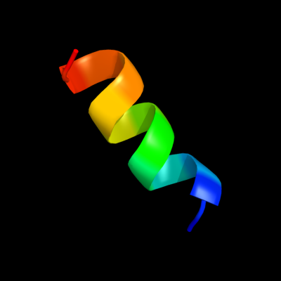
1 c1w5cL_
22.3
54
PDB header: photosynthesisChain: L: PDB Molecule: cytochrome b559 beta subunit;PDBTitle: photosystem ii from thermosynechococcus elongatus
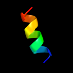
2 d2axtf1
21.8
54
Fold: Single transmembrane helixSuperfamily: Cytochrome b559 subunitsFamily: Cytochrome b559 subunits3 d1c2aa1
19.2
36
Fold: Knottins (small inhibitors, toxins, lectins)Superfamily: Bowman-Birk inhibitor, BBIFamily: Bowman-Birk inhibitor, BBI4 d1fl2a2
15.0
19
Fold: FAD/NAD(P)-binding domainSuperfamily: FAD/NAD(P)-binding domainFamily: FAD/NAD-linked reductases, N-terminal and central domains5 d1j83a_
14.3
38
Fold: Galactose-binding domain-likeSuperfamily: Galactose-binding domain-likeFamily: Family 17 carbohydrate binding module, CBM176 d2fj8a1
13.8
50
Fold: Knottins (small inhibitors, toxins, lectins)Superfamily: Bowman-Birk inhibitor, BBIFamily: Bowman-Birk inhibitor, BBI7 d1efva2
11.1
18
Fold: DHS-like NAD/FAD-binding domainSuperfamily: DHS-like NAD/FAD-binding domainFamily: C-terminal domain of the electron transfer flavoprotein alpha subunit8 c1efpC_
11.0
24
PDB header: electron transportChain: C: PDB Molecule: protein (electron transfer flavoprotein);PDBTitle: electron transfer flavoprotein (etf) from paracoccus2 denitrificans
9 d1efpa2
10.9
18
Fold: DHS-like NAD/FAD-binding domainSuperfamily: DHS-like NAD/FAD-binding domainFamily: C-terminal domain of the electron transfer flavoprotein alpha subunit10 c1tx6J_
10.9
50
PDB header: hydrolase/protein bindingChain: J: PDB Molecule: bowman-birk type trypsin inhibitor;PDBTitle: trypsin:bbi complex
11 c2bi8A_
10.6
39
PDB header: isomeraseChain: A: PDB Molecule: udp-galactopyranose mutase;PDBTitle: udp-galactopyranose mutase from klebsiella pneumoniae with2 reduced fad
12 c1w1nA_
9.5
50
PDB header: transferaseChain: A: PDB Molecule: phosphatidylinositol 3-kinase tor1;PDBTitle: the solution structure of the fatc domain of the protein2 kinase tor1 from yeast
13 d1pbia_
9.4
29
Fold: Knottins (small inhibitors, toxins, lectins)Superfamily: Bowman-Birk inhibitor, BBIFamily: Bowman-Birk inhibitor, BBI14 c2qn5B_
7.6
42
PDB header: hydrolase inhibitor/hydrolaseChain: B: PDB Molecule: bowman-birk type bran trypsin inhibitor;PDBTitle: crystal structure and functional study of the bowman-birk2 inhibitor from rice bran in complex with bovine trypsin
15 c2zylA_
7.4
17
PDB header: oxidoreductaseChain: A: PDB Molecule: possible oxidoreductase;PDBTitle: crystal structure of 3-ketosteroid-9-alpha-hydroxylase2 (ksha) from m. tuberculosis
16 c2pd0D_
7.3
38
PDB header: structural genomics, unknown functionChain: D: PDB Molecule: hypothetical protein;PDBTitle: protein cgd2_2020 from cryptosporidium parvum
17 d3clsd2
7.3
27
Fold: DHS-like NAD/FAD-binding domainSuperfamily: DHS-like NAD/FAD-binding domainFamily: C-terminal domain of the electron transfer flavoprotein alpha subunit18 c2fqcA_
6.9
80
PDB header: toxinChain: A: PDB Molecule: conotoxin pl14a;PDBTitle: solution structure of conotoxin pl14a
19 c1zq1D_
6.7
8
PDB header: lyaseChain: D: PDB Molecule: glutamyl-trna(gln) amidotransferase subunit e;PDBTitle: structure of gatde trna-dependent amidotransferase from2 pyrococcus abyssi
20 d2bi7a1
6.7
37
Fold: Nucleotide-binding domainSuperfamily: Nucleotide-binding domainFamily: UDP-galactopyranose mutase, N-terminal domain21 d2d6fc3
not modelled
6.6
21
Fold: Glutamine synthetase/guanido kinaseSuperfamily: Glutamine synthetase/guanido kinaseFamily: GatB/GatE catalytic domain-like22 d1cqxa2
not modelled
6.2
21
Fold: Reductase/isomerase/elongation factor common domainSuperfamily: Riboflavin synthase domain-likeFamily: Ferredoxin reductase FAD-binding domain-like23 d1u97a_
not modelled
6.1
46
Fold: Cysteine alpha-hairpin motifSuperfamily: Cysteine alpha-hairpin motifFamily: COX17-like24 d1pm6a_
not modelled
5.9
20
Fold: Putative DNA-binding domainSuperfamily: Putative DNA-binding domainFamily: Excisionase-like25 d1qmva_
not modelled
5.8
23
Fold: Thioredoxin foldSuperfamily: Thioredoxin-likeFamily: Glutathione peroxidase-like26 c3fnvB_
not modelled
5.8
40
PDB header: metal binding proteinChain: B: PDB Molecule: cdgsh iron sulfur domain-containing protein 2;PDBTitle: crystal structure of miner1: the redox-active 2fe-2s protein causative2 in wolfram syndrome 2
27 c2pnvA_
not modelled
5.5
50
PDB header: membrane proteinChain: A: PDB Molecule: small conductance calcium-activated potassiumPDBTitle: crystal structure of the leucine zipper domain of small-2 conductance ca2+-activated k+ (skca) channel from rattus3 norvegicus
28 c2rnbA_
not modelled
5.4
54
PDB header: metal transportChain: A: PDB Molecule: cytochrome c oxidase copper chaperone;PDBTitle: solution structure of human cu(i)cox17
29 d2v1ra1
not modelled
5.3
31
Fold: SH3-like barrelSuperfamily: SH3-domainFamily: SH3-domain