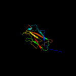
| 1 |
|
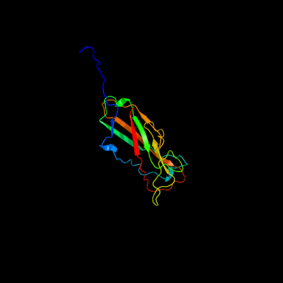
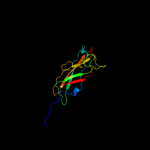
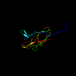
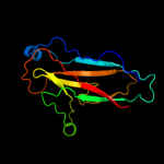
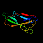
PDB 3jwn chain L
Region: 28 - 179
Aligned: 151
Modelled: 152
Confidence: 100.0%
Identity: 30%
PDB header:protein binding/cell adhesion
Chain: L: PDB Molecule:protein fimf;
PDBTitle: complex of fimc, fimf, fimg and fimh
Phyre2
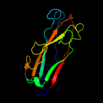
| 2 |
|
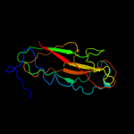
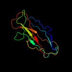
PDB 3jwn chain K
Region: 28 - 179
Aligned: 151
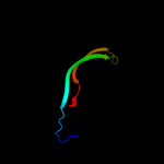
Modelled: 152
Confidence: 100.0%
Identity: 30%
PDB header:protein binding/cell adhesion
Chain: K: PDB Molecule:protein fimf;
PDBTitle: complex of fimc, fimf, fimg and fimh
Phyre2
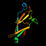
| 3 |
|
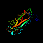
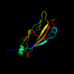
PDB 3jwn chain E
Region: 28 - 179
Aligned: 151
Modelled: 152
Confidence: 100.0%
Identity: 29%
PDB header:protein binding/cell adhesion
Chain: E: PDB Molecule:protein fimf;
PDBTitle: complex of fimc, fimf, fimg and fimh
Phyre2
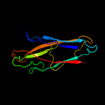
| 4 |
|
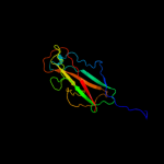
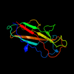
PDB 3jwn chain F
Region: 28 - 179
Aligned: 151
Modelled: 152
Confidence: 100.0%
Identity: 29%
PDB header:protein binding/cell adhesion
Chain: F: PDB Molecule:protein fimf;
PDBTitle: complex of fimc, fimf, fimg and fimh
Phyre2
| 5 |
|
PDB 2jmr chain A
Region: 27 - 179
Aligned: 152
Modelled: 153
Confidence: 100.0%
Identity: 30%
PDB header:cell adhesion
Chain: A: PDB Molecule:fimf;
PDBTitle: nmr structure of the e. coli type 1 pilus subunit fimf
Phyre2
| 6 |
|
PDB 2jty chain A
Region: 23 - 179
Aligned: 156
Modelled: 157
Confidence: 100.0%
Identity: 33%
PDB header:structural protein
Chain: A: PDB Molecule:type-1 fimbrial protein, a chain;
PDBTitle: self-complemented variant of fima, the main subunit of type 1 pilus
Phyre2
| 7 |
|
PDB 2j2z chain B domain 1
Region: 31 - 179
Aligned: 143
Modelled: 149
Confidence: 99.9%
Identity: 20%
Fold: Common fold of diphtheria toxin/transcription factors/cytochrome f
Superfamily: Bacterial adhesins
Family: Pilus subunits
Phyre2
| 8 |
|
PDB 2uy6 chain B domain 1
Region: 31 - 179
Aligned: 143
Modelled: 149
Confidence: 99.9%
Identity: 27%
Fold: Common fold of diphtheria toxin/transcription factors/cytochrome f
Superfamily: Bacterial adhesins
Family: Pilus subunits
Phyre2
| 9 |
|
PDB 1pdk chain B
Region: 38 - 179
Aligned: 140
Modelled: 142
Confidence: 99.9%
Identity: 26%
Fold: Common fold of diphtheria toxin/transcription factors/cytochrome f
Superfamily: Bacterial adhesins
Family: Pilus subunits
Phyre2
| 10 |
|
PDB 3bfw chain A
Region: 40 - 178
Aligned: 131
Modelled: 139
Confidence: 99.9%
Identity: 26%
PDB header:structural protein/structural protein
Chain: A: PDB Molecule:protein fimg;
PDBTitle: crystal structure of truncated fimg (fimgt) in complex with the donor2 strand peptide of fimf (dsf)
Phyre2
| 11 |
|
PDB 3bwu chain F
Region: 58 - 179
Aligned: 121
Modelled: 122
Confidence: 99.9%
Identity: 33%
PDB header:chaperone, structural, membrane protein
Chain: F: PDB Molecule:protein fimf;
PDBTitle: crystal structure of the ternary complex of fimd (n-terminal domain,2 fimdn) with fimc and the n-terminally truncated pilus subunit fimf3 (fimft)
Phyre2
| 12 |
|
PDB 1klf chain P
Region: 31 - 179
Aligned: 133
Modelled: 146
Confidence: 99.8%
Identity: 21%
PDB header:chaperone/adhesin complex
Chain: P: PDB Molecule:fimh protein;
PDBTitle: fimh adhesin-fimc chaperone complex with d-mannose
Phyre2
| 13 |
|
PDB 1ze3 chain H domain 1
Region: 41 - 179
Aligned: 121
Modelled: 127
Confidence: 99.8%
Identity: 22%
Fold: Common fold of diphtheria toxin/transcription factors/cytochrome f
Superfamily: Bacterial adhesins
Family: Pilus subunits
Phyre2
| 14 |
|
PDB 2w07 chain B
Region: 37 - 178
Aligned: 119
Modelled: 142
Confidence: 99.8%
Identity: 19%
PDB header:cell adhesion
Chain: B: PDB Molecule:minor pilin subunit papf;
PDBTitle: structural determinants of polymerization reactivity of the2 p pilus adaptor subunit papf
Phyre2
| 15 |
|
PDB 1n12 chain A
Region: 41 - 178
Aligned: 129
Modelled: 138
Confidence: 99.6%
Identity: 14%
Fold: Common fold of diphtheria toxin/transcription factors/cytochrome f
Superfamily: Bacterial adhesins
Family: Pilus subunits
Phyre2
| 16 |
|
PDB 2wmp chain B
Region: 45 - 178
Aligned: 116
Modelled: 134
Confidence: 84.3%
Identity: 12%
PDB header:chaperone
Chain: B: PDB Molecule:papg protein;
PDBTitle: structure of the e. coli chaperone papd in complex with the pilin2 domain of the papgii adhesin
Phyre2
| 17 |
|
PDB 1p5v chain B
Region: 77 - 179
Aligned: 100
Modelled: 103
Confidence: 33.9%
Identity: 15%
Fold: Common fold of diphtheria toxin/transcription factors/cytochrome f
Superfamily: Bacterial adhesins
Family: Pilus subunits
Phyre2
| 18 |
|
PDB 1w3g chain A
Region: 130 - 179
Aligned: 50
Modelled: 50
Confidence: 9.3%
Identity: 16%
PDB header:toxin/lectin
Chain: A: PDB Molecule:hemolytic lectin from laetiporus sulphureus;
PDBTitle: hemolytic lectin from the mushroom laetiporus sulphureus2 complexed with two n-acetyllactosamine molecules.
Phyre2