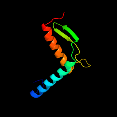
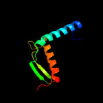
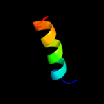
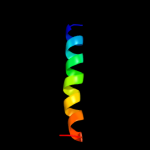
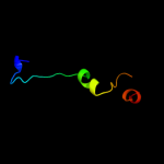
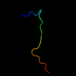
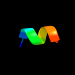
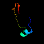
PDB header:structural genomics, unknown function Chain: B: PDB Molecule:protein ycin; PDBTitle: the structure of ycin, an unchracterized protein from shigella2 flexneri.
Confidence and coverage
Confidence:
100.0%
Coverage:
98%
81 residues ( 98% of your sequence) have been modelled with 100.0% confidence by the single highest scoring template.
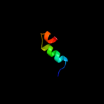
Region: 26 - 44 Aligned: 19 Modelled: 19 Confidence: 5.4% Identity: 16% PDB header:ribosome Chain: L: PDB Molecule:40s ribosomal protein s23; PDBTitle: structure of the ribosomal 80s-eef2-sordarin complex from2 yeast obtained by docking atomic models for rna and protein3 components into a 11.7 a cryo-em map. this file, 1s1h,4 contains 40s subunit. the 60s ribosomal subunit is in file5 1s1i.
Phyre2
21
22
23
24
Detailed template information
Binding site prediction
Due to computational demand, binding site predictions are not run for batch jobs
If you want to predict binding sites, please manually submit your model of choice to 3DLigandSite
Phyre is for academic use only
Please cite: Protein structure prediction on
the web: a case study using the Phyre server
Kelley LA and Sternberg MJE. Nature Protocols
4, 363 - 371 (2009) [pdf] [Import into BibTeX]
If you use the binding site
predictions from 3DLigandSite, please also cite:
3DLigandSite: predicting ligand-binding sites using similar structures.
Wass MN, Kelley LA and Sternberg
MJ Nucleic Acids Research 38, W469-73 (2010) [PubMed]