Phyre Home Retrieve Phyre Job Id
Emaill.a.kelley@imperial.ac.uk
DescriptionP65294
DateThu Jan 5 12:10:11 GMT 2012
Unique Job IDca51ccaca33865ce

Summary 

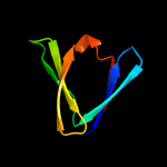
Top model
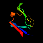
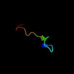
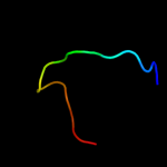
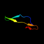
Image coloured by rainbow N → C terminus
Model (left) based on template d2rb6a1
Top template information
Fold:Sm-like fold
Superfamily:Sm-like ribonucleoproteins
Family:YgdI/YgdR-like
Confidence and coverage
Confidence: 99.9% Coverage: 72%
52 residues ( 72% of your sequence) have been modelled with 99.9% confidence by the single highest scoring template.
You may wish to submit your sequence to Phyrealarm. This will automatically scan your sequence every week for new potential templates as they appear in the Phyre2 library.
3D viewing
Interactive 3D view in Jmol

Sequence analysis 

Secondary structure and disorder prediction 

   1........10.........20.........30.........40.........50.........60
Sequence  MKKWAVIISAVGLAFAVSGCSSDYVMATKDGRMILTDGKPEIDDDTGLVSYHDQQGNAMQ
Secondary structure 


SS confidence 


Disorder  ????


???


??


Disorder confidence 


   .........70..
Sequence  INRDDVSQIIER
Secondary structure 

SS confidence 


Disorder 
?


????
Disorder confidence 


Confidence Key
High(9)                    Low (0)
?Disordered
Alpha helix
Beta strand

Domain analysis 

Hover over an aligned region to see model and summary info

Please note, only up to the top 20 hits are modelled to reduce computer load

RankAligned region

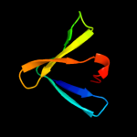
PDB 2rb6 chain A domain 1

3D model

Region: 21 - 72
Aligned: 52
Modelled: 52
Confidence: 99.9%
Identity: 48%
Fold: Sm-like fold
Superfamily: Sm-like ribonucleoproteins
Family: YgdI/YgdR-like

Phyre2

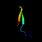
PDB 2k57 chain A domain 1

3D model

Region: 21 - 71
Aligned: 51
Modelled: 51
Confidence: 99.9%
Identity: 27%
Fold: Sm-like fold
Superfamily: Sm-like ribonucleoproteins
Family: YgdI/YgdR-like

Phyre2

PDB 3bdu chain A domain 1

3D model

Region: 21 - 71
Aligned: 51
Modelled: 51
Confidence: 99.9%
Identity: 51%
Fold: Sm-like fold
Superfamily: Sm-like ribonucleoproteins
Family: YgdI/YgdR-like

Phyre2

PDB 2jn0 chain A domain 1

3D model

Region: 23 - 72
Aligned: 50
Modelled: 50
Confidence: 99.9%
Identity: 100%
Fold: Sm-like fold
Superfamily: Sm-like ribonucleoproteins
Family: YgdI/YgdR-like

Phyre2

PDB 2rd1 chain A domain 1

3D model

Region: 22 - 71
Aligned: 50
Modelled: 50
Confidence: 99.9%
Identity: 50%
Fold: Sm-like fold
Superfamily: Sm-like ribonucleoproteins
Family: YgdI/YgdR-like

Phyre2

PDB 2ra2 chain A domain 1

3D model

Region: 22 - 71
Aligned: 50
Modelled: 47
Confidence: 99.9%
Identity: 54%
Fold: Sm-like fold
Superfamily: Sm-like ribonucleoproteins
Family: YgdI/YgdR-like

Phyre2

PDB 2npt chain A domain 1

3D model

Region: 43 - 65
Aligned: 23
Modelled: 23
Confidence: 45.3%
Identity: 22%
Fold: beta-Grasp (ubiquitin-like)
Superfamily: CAD & PB1 domains
Family: PB1 domain

Phyre2

PDB 1wi0 chain A

3D model

Region: 43 - 65
Aligned: 23
Modelled: 23
Confidence: 35.6%
Identity: 22%
Fold: beta-Grasp (ubiquitin-like)
Superfamily: CAD & PB1 domains
Family: PB1 domain

Phyre2

PDB 2h2w chain A

3D model

Region: 24 - 46
Aligned: 23
Modelled: 23
Confidence: 24.6%
Identity: 48%
PDB header:transferase
Chain: A: PDB Molecule:homoserine o-succinyltransferase;
PDBTitle: crystal structure of homoserine o-succinyltransferase (ec 2.3.1.46)2 (homoserine o-transsuccinylase) (hts) (tm0881) from thermotoga3 maritima at 2.52 a resolution

Phyre2

PDB 3dso chain A

3D model

Region: 25 - 38
Aligned: 14
Modelled: 14
Confidence: 23.8%
Identity: 50%
PDB header:metal binding protein
Chain: A: PDB Molecule:putative uncharacterized protein copk;
PDBTitle: crystal structure of cu(i) bound copper resistance protein copk

Phyre2

PDB 2ghr chain A domain 1

3D model

Region: 23 - 46
Aligned: 24
Modelled: 24
Confidence: 22.6%
Identity: 25%
Fold: Flavodoxin-like
Superfamily: Class I glutamine amidotransferase-like
Family: HTS-like

Phyre2

PDB 2ktr chain A

3D model

Region: 40 - 65
Aligned: 26
Modelled: 26
Confidence: 20.1%
Identity: 15%
PDB header:signaling protein, transport protein
Chain: A: PDB Molecule:sequestosome-1;
PDBTitle: nmr structure of p62 pb1 dimer determined based on pcs

Phyre2

PDB 1br2 chain A domain 1

3D model

Region: 54 - 69
Aligned: 16
Modelled: 16
Confidence: 16.6%
Identity: 19%
Fold: SH3-like barrel
Superfamily: Myosin S1 fragment, N-terminal domain
Family: Myosin S1 fragment, N-terminal domain

Phyre2

PDB 2kdk chain A

3D model

Region: 24 - 58
Aligned: 35
Modelled: 35
Confidence: 15.6%
Identity: 23%
PDB header:transcription regulator
Chain: A: PDB Molecule:aryl hydrocarbon receptor nuclear translocator-like protein
PDBTitle: structure of human circadian clock protein bmal2 c-terminal pas domain

Phyre2

PDB 1gvi chain A domain 2

3D model

Region: 43 - 65
Aligned: 23
Modelled: 23
Confidence: 15.2%
Identity: 39%
Fold: Glycosyl hydrolase domain
Superfamily: Glycosyl hydrolase domain
Family: alpha-Amylases, C-terminal beta-sheet domain

Phyre2

PDB 1h6l chain A

3D model

Region: 43 - 60
Aligned: 17
Modelled: 18
Confidence: 11.5%
Identity: 24%
Fold: 6-bladed beta-propeller
Superfamily: Thermostable phytase (3-phytase)
Family: Thermostable phytase (3-phytase)

Phyre2

PDB 2k5w chain A domain 1

3D model

Region: 33 - 61
Aligned: 29
Modelled: 29
Confidence: 9.4%
Identity: 34%
Fold: OB-fold
Superfamily: BC4932-like
Family: BC4932-like

Phyre2

PDB 2pxg chain A

3D model

Region: 17 - 31
Aligned: 15
Modelled: 15
Confidence: 9.3%
Identity: 20%
PDB header:membrane protein
Chain: A: PDB Molecule:outer membrane protein;
PDBTitle: nmr solution structure of omla

Phyre2

PDB 1w0n chain A

3D model

Region: 15 - 30
Aligned: 16
Modelled: 16
Confidence: 9.1%
Identity: 38%
Fold: Galactose-binding domain-like
Superfamily: Galactose-binding domain-like
Family: Family 36 carbohydrate binding module, CBM36

Phyre2

PDB 2b38 chain A

3D model

Region: 18 - 32
Aligned: 15
Modelled: 15
Confidence: 9.0%
Identity: 47%
PDB header:plant protein
Chain: A: PDB Molecule:kalata b8;
PDBTitle: solution structure of kalata b8

Phyre2
1

d2rb6a1
2

d2k57a1
3

d3bdua1
4

d2jn0a1
5

d2rd1a1
6

d2ra2a1
7

d2npta1
8

d1wi0a_
9

c2h2wA_
10

c3dsoA_
11

d2ghra1
12

c2ktrA_
13

d1br2a1
14

c2kdkA_
15

d1gvia2
16

d1h6la_
17

d2k5wa1
18

c2pxgA_
19

d1w0na_
20

c2b38A_
21


22


23


24


25


26


27


28


Detailed template information 

#
Template Alignment Coverage3D Model Confidence
% i.d. Template Information
1d2rb6a1


99.9 48 Fold:Sm-like fold
Superfamily:Sm-like ribonucleoproteins
Family:YgdI/YgdR-like
2d2k57a1


99.9 27 Fold:Sm-like fold
Superfamily:Sm-like ribonucleoproteins
Family:YgdI/YgdR-like
3d3bdua1


99.9 51 Fold:Sm-like fold
Superfamily:Sm-like ribonucleoproteins
Family:YgdI/YgdR-like
4d2jn0a1


99.9 100 Fold:Sm-like fold
Superfamily:Sm-like ribonucleoproteins
Family:YgdI/YgdR-like
5d2rd1a1


99.9 50 Fold:Sm-like fold
Superfamily:Sm-like ribonucleoproteins
Family:YgdI/YgdR-like
6d2ra2a1


99.9 54 Fold:Sm-like fold
Superfamily:Sm-like ribonucleoproteins
Family:YgdI/YgdR-like
7d2npta1


45.3 22 Fold:beta-Grasp (ubiquitin-like)
Superfamily:CAD & PB1 domains
Family:PB1 domain
8d1wi0a_


35.6 22 Fold:beta-Grasp (ubiquitin-like)
Superfamily:CAD & PB1 domains
Family:PB1 domain
9c2h2wA_


24.6 48 PDB header:transferase
Chain: A: PDB Molecule:homoserine o-succinyltransferase;
PDBTitle: crystal structure of homoserine o-succinyltransferase (ec 2.3.1.46)2 (homoserine o-transsuccinylase) (hts) (tm0881) from thermotoga3 maritima at 2.52 a resolution
10c3dsoA_


23.8 50 PDB header:metal binding protein
Chain: A: PDB Molecule:putative uncharacterized protein copk;
PDBTitle: crystal structure of cu(i) bound copper resistance protein copk
11d2ghra1


22.6 25 Fold:Flavodoxin-like
Superfamily:Class I glutamine amidotransferase-like
Family:HTS-like
12c2ktrA_


20.1 15 PDB header:signaling protein, transport protein
Chain: A: PDB Molecule:sequestosome-1;
PDBTitle: nmr structure of p62 pb1 dimer determined based on pcs
13d1br2a1


16.6 19 Fold:SH3-like barrel
Superfamily:Myosin S1 fragment, N-terminal domain
Family:Myosin S1 fragment, N-terminal domain
14c2kdkA_


15.6 23 PDB header:transcription regulator
Chain: A: PDB Molecule:aryl hydrocarbon receptor nuclear translocator-like protein
PDBTitle: structure of human circadian clock protein bmal2 c-terminal pas domain
15d1gvia2


15.2 39 Fold:Glycosyl hydrolase domain
Superfamily:Glycosyl hydrolase domain
Family:alpha-Amylases, C-terminal beta-sheet domain
16d1h6la_


11.5 24 Fold:6-bladed beta-propeller
Superfamily:Thermostable phytase (3-phytase)
Family:Thermostable phytase (3-phytase)
17d2k5wa1


9.4 34 Fold:OB-fold
Superfamily:BC4932-like
Family:BC4932-like
18c2pxgA_


9.3 20 PDB header:membrane protein
Chain: A: PDB Molecule:outer membrane protein;
PDBTitle: nmr solution structure of omla
19d1w0na_


9.1 38 Fold:Galactose-binding domain-like
Superfamily:Galactose-binding domain-like
Family:Family 36 carbohydrate binding module, CBM36
20c2b38A_


9.0 47 PDB header:plant protein
Chain: A: PDB Molecule:kalata b8;
PDBTitle: solution structure of kalata b8
21d1wmha_


not modelled 8.1 12 Fold:beta-Grasp (ubiquitin-like)
Superfamily:CAD & PB1 domains
Family:PB1 domain
22d1wzaa1


not modelled 7.0 23 Fold:Glycosyl hydrolase domain
Superfamily:Glycosyl hydrolase domain
Family:alpha-Amylases, C-terminal beta-sheet domain
23d2bkfa1


not modelled 6.9 24 Fold:beta-Grasp (ubiquitin-like)
Superfamily:CAD & PB1 domains
Family:PB1 domain
24d1u5sa1


not modelled 6.6 11 Fold:SH3-like barrel
Superfamily:SH3-domain
Family:SH3-domain
25d1wj6a_


not modelled 6.6 24 Fold:beta-Grasp (ubiquitin-like)
Superfamily:CAD & PB1 domains
Family:PB1 domain
26d1sg5a1


not modelled 5.9 11 Fold:Rof/RNase P subunit-like
Superfamily:Rof/RNase P subunit-like
Family:Rof-like
27c1tr8A_


not modelled 5.5 21 PDB header:chaperone
Chain: A: PDB Molecule:conserved protein (mth177);
PDBTitle: crystal structure of archaeal nascent polypeptide-associated complex2 (aenac)
28c3uc0B_


not modelled 5.3 36 PDB header:viral protein/immune system
Chain: B: PDB Molecule:envelope protein;
PDBTitle: crystal structure of domain i of the envelope glycoprotein ectodomain2 from dengue virus serotype 4 in complex with the fab fragment of the3 chimpanzee monoclonal antibody 5h2

Binding site prediction 

Due to computational demand, binding site predictions are not run for batch jobs

If you want to predict binding sites, please manually submit your model of choice to 3DLigandSite


Phyre is for academic use only

Please cite: Protein structure prediction on the web: a case study using the Phyre server
Kelley LA and Sternberg MJE. Nature Protocols 4, 363 - 371 (2009) [pdf] [Import into BibTeX]
 
If you use the binding site predictions from 3DLigandSite, please also cite:
3DLigandSite: predicting ligand-binding sites using similar structures.
Wass MN, Kelley LA and Sternberg MJ Nucleic Acids Research 38, W469-73 (2010) [PubMed]
 
© Structural Bioinformatics Group
Imperial College London
Lawrence Kelley, Benjamin Jefferys
Disclaimer
Terms and Conditions
Component software
Template detection: HHpred 1.51
Secondary structure prediction: Psi-pred 2.5
Disorder prediction: Disopred 2.4
Transmembrane prediction: Memsat_SVM
Multi-template modelling and ab initio: Poing 1.0