Phyre Home Retrieve Phyre Job Id
Emaill.a.kelley@imperial.ac.uk
DescriptionP76154
DateThu Jan 5 12:19:46 GMT 2012
Unique Job IDc691a432ba458a37

Summary 

Top model
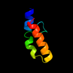
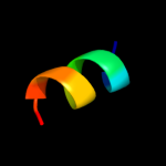
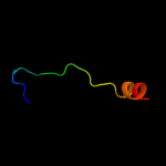
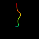
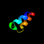
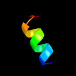
Image coloured by rainbow N → C terminus
Model (left) based on template c2kk1A_
Top template information
PDB header:transferase
Chain: A: PDB Molecule:tyrosine-protein kinase abl2;
PDBTitle: solution structure of c-terminal domain of tyrosine-protein2 kinase abl2 from homo sapiens, northeast structural3 genomics consortium (nesg) target hr5537a
Confidence and coverage
Confidence: 36.9% Coverage: 51%
39 residues ( 51% of your sequence) have been modelled with 36.9% confidence by the single highest scoring template.
You may wish to submit your sequence to Phyrealarm. This will automatically scan your sequence every week for new potential templates as they appear in the Phyre2 library.
Please note: You must be registered and logged in to use Phyrealarm.
3D viewing
Interactive 3D view in Jmol

Sequence analysis 

Secondary structure and disorder prediction 

   1........10.........20.........30.........40.........50.........60
Sequence  MKSKDTLKWFPAQLPEVRIILGDAVVEVAKQGRPINTRTLLDYIEGNIKKKSWLDNKELL
Secondary structure 


SS confidence 


Disorder  ???????


?
?


??


???


Disorder confidence 


   .........70.......
Sequence  QTAISVLKDNQNLNGKM
Secondary structure 


SS confidence 


Disorder 


??
???????
Disorder confidence 


Confidence Key
High(9)                    Low (0)
?Disordered
Alpha helix
Beta strand

Domain analysis 

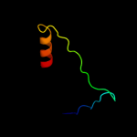
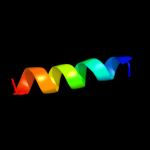
Hover over an aligned region to see model and summary info

Please note, only up to the top 20 hits are modelled to reduce computer load

RankAligned region

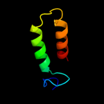
PDB 2kk1 chain A

3D model

Region: 24 - 70
Aligned: 39
Modelled: 47
Confidence: 36.9%
Identity: 31%
PDB header:transferase
Chain: A: PDB Molecule:tyrosine-protein kinase abl2;
PDBTitle: solution structure of c-terminal domain of tyrosine-protein2 kinase abl2 from homo sapiens, northeast structural3 genomics consortium (nesg) target hr5537a

Phyre2

PDB 1zzp chain A

3D model

Region: 24 - 70
Aligned: 39
Modelled: 47
Confidence: 25.0%
Identity: 31%
PDB header:transferase
Chain: A: PDB Molecule:proto-oncogene tyrosine-protein kinase abl1;
PDBTitle: solution structure of the f-actin binding domain of bcr-2 abl/c-abl

Phyre2

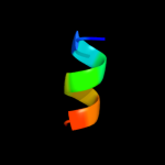
PDB 1a11 chain A

3D model

Region: 58 - 67
Aligned: 10
Modelled: 10
Confidence: 24.1%
Identity: 70%
PDB header:acetylcholine receptor
Chain: A: PDB Molecule:acetylcholine receptor m2;
PDBTitle: nmr structure of membrane spanning segment 2 of the2 acetylcholine receptor in dpc micelles, 10 structures

Phyre2

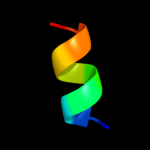
PDB 1eq8 chain C

3D model

Region: 58 - 67
Aligned: 10
Modelled: 10
Confidence: 19.7%
Identity: 70%
PDB header:signaling protein
Chain: C: PDB Molecule:acetylcholine receptor protein;
PDBTitle: three-dimensional structure of the pentameric helical2 bundle of the acetylcholine receptor m2 transmembrane3 segment

Phyre2

PDB 1eq8 chain D

3D model

Region: 58 - 67
Aligned: 10
Modelled: 10
Confidence: 19.7%
Identity: 70%
PDB header:signaling protein
Chain: D: PDB Molecule:acetylcholine receptor protein;
PDBTitle: three-dimensional structure of the pentameric helical2 bundle of the acetylcholine receptor m2 transmembrane3 segment

Phyre2

PDB 1eq8 chain A

3D model

Region: 58 - 67
Aligned: 10
Modelled: 10
Confidence: 19.7%
Identity: 70%
PDB header:signaling protein
Chain: A: PDB Molecule:acetylcholine receptor protein;
PDBTitle: three-dimensional structure of the pentameric helical2 bundle of the acetylcholine receptor m2 transmembrane3 segment

Phyre2

PDB 1eq8 chain E

3D model

Region: 58 - 67
Aligned: 10
Modelled: 10
Confidence: 19.7%
Identity: 70%
PDB header:signaling protein
Chain: E: PDB Molecule:acetylcholine receptor protein;
PDBTitle: three-dimensional structure of the pentameric helical2 bundle of the acetylcholine receptor m2 transmembrane3 segment

Phyre2

PDB 1eq8 chain B

3D model

Region: 58 - 67
Aligned: 10
Modelled: 10
Confidence: 19.7%
Identity: 70%
PDB header:signaling protein
Chain: B: PDB Molecule:acetylcholine receptor protein;
PDBTitle: three-dimensional structure of the pentameric helical2 bundle of the acetylcholine receptor m2 transmembrane3 segment

Phyre2

PDB 1wq6 chain A

3D model

Region: 36 - 72
Aligned: 28
Modelled: 37
Confidence: 17.5%
Identity: 25%
PDB header:oncoprotein
Chain: A: PDB Molecule:aml1-eto;
PDBTitle: the tetramer structure of the nervy homolgy two (nhr2) domain of aml1-2 eto is critical for aml1-eto's activity

Phyre2

PDB 1m5q chain 1

3D model

Region: 19 - 45
Aligned: 27
Modelled: 27
Confidence: 11.0%
Identity: 22%
Fold: Sm-like fold
Superfamily: Sm-like ribonucleoproteins
Family: Sm motif of small nuclear ribonucleoproteins, SNRNP

Phyre2

PDB 2fna chain A domain 1

3D model

Region: 29 - 49
Aligned: 21
Modelled: 21
Confidence: 9.4%
Identity: 19%
Fold: DNA/RNA-binding 3-helical bundle
Superfamily: "Winged helix" DNA-binding domain
Family: Helicase DNA-binding domain

Phyre2

PDB 3e0z chain B

3D model

Region: 35 - 58
Aligned: 24
Modelled: 23
Confidence: 7.3%
Identity: 29%
PDB header:unknown function
Chain: B: PDB Molecule:protein of unknown function;
PDBTitle: crystal structure of a putative imidazole glycerol phosphate synthase2 homolog (eubrec_1070) from eubacterium rectale at 1.75 a resolution

Phyre2

PDB 3nbm chain A

3D model

Region: 15 - 55
Aligned: 41
Modelled: 41
Confidence: 6.2%
Identity: 22%
PDB header:transferase
Chain: A: PDB Molecule:pts system, lactose-specific iibc components;
PDBTitle: the lactose-specific iib component domain structure of the2 phosphoenolpyruvate:carbohydrate phosphotransferase system (pts) from3 streptococcus pneumoniae.

Phyre2

PDB 1izl chain J

3D model

Region: 31 - 37
Aligned: 7
Modelled: 7
Confidence: 6.2%
Identity: 57%
PDB header:photosynthesis
Chain: J: PDB Molecule:photosystem ii: subunit psba;
PDBTitle: crystal structure of photosystem ii

Phyre2

PDB 3blh chain B domain 1

3D model

Region: 38 - 69
Aligned: 26
Modelled: 32
Confidence: 5.7%
Identity: 15%
Fold: Cyclin-like
Superfamily: Cyclin-like
Family: Cyclin

Phyre2

PDB 1mu2 chain A

3D model

Region: 12 - 50
Aligned: 38
Modelled: 39
Confidence: 5.7%
Identity: 16%
PDB header:transferase
Chain: A: PDB Molecule:hiv-2 rt;
PDBTitle: crystal structure of hiv-2 reverse transcriptase

Phyre2

PDB 3ctd chain B

3D model

Region: 14 - 29
Aligned: 16
Modelled: 16
Confidence: 5.7%
Identity: 31%
PDB header:structural genomics, unknown function
Chain: B: PDB Molecule:putative atpase, aaa family;
PDBTitle: crystal structure of a putative aaa family atpase from2 prochlorococcus marinus subsp. pastoris

Phyre2

PDB 1dfw chain A

3D model

Region: 38 - 50
Aligned: 13
Modelled: 13
Confidence: 5.7%
Identity: 38%
PDB header:immune system
Chain: A: PDB Molecule:lung surfactant protein b;
PDBTitle: conformational mapping of the n-terminal segment of2 surfactant protein b in lipid using 13c-enhanced fourier3 transform infrared spectroscopy (ftir)

Phyre2

PDB 1wn4 chain A

3D model

Region: 20 - 32
Aligned: 13
Modelled: 13
Confidence: 5.5%
Identity: 46%
PDB header:plant protein
Chain: A: PDB Molecule:vontr protein;
PDBTitle: nmr structure of vontr

Phyre2

PDB 2r5u chain D

3D model

Region: 23 - 47
Aligned: 25
Modelled: 25
Confidence: 5.4%
Identity: 20%
PDB header:hydrolase
Chain: D: PDB Molecule:replicative dna helicase;
PDBTitle: crystal structure of the n-terminal domain of dnab helicase from2 mycobacterium tuberculosis

Phyre2
1

c2kk1A_
2

c1zzpA_
3

c1a11A_
4

c1eq8C_
5

c1eq8D_
6

c1eq8A_
7

c1eq8E_
8

c1eq8B_
9

c1wq6A_
10

d1m5q1_
11

d2fnaa1
12

c3e0zB_
13

c3nbmA_
14

c1izlJ_
15

d3blhb1
16

c1mu2A_
17

c3ctdB_
18

c1dfwA_
19

c1wn4A_
20

c2r5uD_
21


22


23


24


25


26


27


Detailed template information 

#
Template Alignment Coverage3D Model Confidence
% i.d. Template Information
1c2kk1A_


36.9 31 PDB header:transferase
Chain: A: PDB Molecule:tyrosine-protein kinase abl2;
PDBTitle: solution structure of c-terminal domain of tyrosine-protein2 kinase abl2 from homo sapiens, northeast structural3 genomics consortium (nesg) target hr5537a
2c1zzpA_


25.0 31 PDB header:transferase
Chain: A: PDB Molecule:proto-oncogene tyrosine-protein kinase abl1;
PDBTitle: solution structure of the f-actin binding domain of bcr-2 abl/c-abl
3c1a11A_


24.1 70 PDB header:acetylcholine receptor
Chain: A: PDB Molecule:acetylcholine receptor m2;
PDBTitle: nmr structure of membrane spanning segment 2 of the2 acetylcholine receptor in dpc micelles, 10 structures
4c1eq8C_


19.7 70 PDB header:signaling protein
Chain: C: PDB Molecule:acetylcholine receptor protein;
PDBTitle: three-dimensional structure of the pentameric helical2 bundle of the acetylcholine receptor m2 transmembrane3 segment
5c1eq8D_


19.7 70 PDB header:signaling protein
Chain: D: PDB Molecule:acetylcholine receptor protein;
PDBTitle: three-dimensional structure of the pentameric helical2 bundle of the acetylcholine receptor m2 transmembrane3 segment
6c1eq8A_


19.7 70 PDB header:signaling protein
Chain: A: PDB Molecule:acetylcholine receptor protein;
PDBTitle: three-dimensional structure of the pentameric helical2 bundle of the acetylcholine receptor m2 transmembrane3 segment
7c1eq8E_


19.7 70 PDB header:signaling protein
Chain: E: PDB Molecule:acetylcholine receptor protein;
PDBTitle: three-dimensional structure of the pentameric helical2 bundle of the acetylcholine receptor m2 transmembrane3 segment
8c1eq8B_


19.7 70 PDB header:signaling protein
Chain: B: PDB Molecule:acetylcholine receptor protein;
PDBTitle: three-dimensional structure of the pentameric helical2 bundle of the acetylcholine receptor m2 transmembrane3 segment
9c1wq6A_


17.5 25 PDB header:oncoprotein
Chain: A: PDB Molecule:aml1-eto;
PDBTitle: the tetramer structure of the nervy homolgy two (nhr2) domain of aml1-2 eto is critical for aml1-eto's activity
10d1m5q1_


11.0 22 Fold:Sm-like fold
Superfamily:Sm-like ribonucleoproteins
Family:Sm motif of small nuclear ribonucleoproteins, SNRNP
11d2fnaa1


9.4 19 Fold:DNA/RNA-binding 3-helical bundle
Superfamily:"Winged helix" DNA-binding domain
Family:Helicase DNA-binding domain
12c3e0zB_


7.3 29 PDB header:unknown function
Chain: B: PDB Molecule:protein of unknown function;
PDBTitle: crystal structure of a putative imidazole glycerol phosphate synthase2 homolog (eubrec_1070) from eubacterium rectale at 1.75 a resolution
13c3nbmA_


6.2 22 PDB header:transferase
Chain: A: PDB Molecule:pts system, lactose-specific iibc components;
PDBTitle: the lactose-specific iib component domain structure of the2 phosphoenolpyruvate:carbohydrate phosphotransferase system (pts) from3 streptococcus pneumoniae.
14c1izlJ_


6.2 57 PDB header:photosynthesis
Chain: J: PDB Molecule:photosystem ii: subunit psba;
PDBTitle: crystal structure of photosystem ii
15d3blhb1


5.7 15 Fold:Cyclin-like
Superfamily:Cyclin-like
Family:Cyclin
16c1mu2A_


5.7 16 PDB header:transferase
Chain: A: PDB Molecule:hiv-2 rt;
PDBTitle: crystal structure of hiv-2 reverse transcriptase
17c3ctdB_


5.7 31 PDB header:structural genomics, unknown function
Chain: B: PDB Molecule:putative atpase, aaa family;
PDBTitle: crystal structure of a putative aaa family atpase from2 prochlorococcus marinus subsp. pastoris
18c1dfwA_


5.7 38 PDB header:immune system
Chain: A: PDB Molecule:lung surfactant protein b;
PDBTitle: conformational mapping of the n-terminal segment of2 surfactant protein b in lipid using 13c-enhanced fourier3 transform infrared spectroscopy (ftir)
19c1wn4A_


5.5 46 PDB header:plant protein
Chain: A: PDB Molecule:vontr protein;
PDBTitle: nmr structure of vontr
20c2r5uD_


5.4 20 PDB header:hydrolase
Chain: D: PDB Molecule:replicative dna helicase;
PDBTitle: crystal structure of the n-terminal domain of dnab helicase from2 mycobacterium tuberculosis
21d2ivxa2


not modelled 5.3 15 Fold:Cyclin-like
Superfamily:Cyclin-like
Family:Cyclin
22d1b79a_


not modelled 5.2 26 Fold:N-terminal domain of DnaB helicase
Superfamily:N-terminal domain of DnaB helicase
Family:N-terminal domain of DnaB helicase
23c2k37A_


not modelled 5.2 14 PDB header:photosynthesis
Chain: A: PDB Molecule:chlorosome protein a;
PDBTitle: csma
24d2axta1


not modelled 5.1 57 Fold:Bacterial photosystem II reaction centre, L and M subunits
Superfamily:Bacterial photosystem II reaction centre, L and M subunits
Family:Bacterial photosystem II reaction centre, L and M subunits
25c3k13A_


not modelled 5.1 18 PDB header:transferase
Chain: A: PDB Molecule:5-methyltetrahydrofolate-homocysteine methyltransferase;
PDBTitle: structure of the pterin-binding domain metr of 5-2 methyltetrahydrofolate-homocysteine methyltransferase from3 bacteroides thetaiotaomicron
26d3ctda1


not modelled 5.1 31 Fold:post-AAA+ oligomerization domain-like
Superfamily:post-AAA+ oligomerization domain-like
Family:MgsA/YrvN C-terminal domain-like
27c1cekA_


not modelled 5.0 100 PDB header:acetylcholine receptor
Chain: A: PDB Molecule:protein (acetylcholine receptor m2);
PDBTitle: three-dimensional structure of the membrane-embedded m22 channel-lining segment from the nicotinic acetylcholine3 receptor by solid-state nmr spectroscopy

Binding site prediction 

Due to computational demand, binding site predictions are not run for batch jobs

If you want to predict binding sites, please manually submit your model of choice to 3DLigandSite


Phyre is for academic use only

Please cite: Protein structure prediction on the web: a case study using the Phyre server
Kelley LA and Sternberg MJE. Nature Protocols 4, 363 - 371 (2009) [pdf] [Import into BibTeX]
 
If you use the binding site predictions from 3DLigandSite, please also cite:
3DLigandSite: predicting ligand-binding sites using similar structures.
Wass MN, Kelley LA and Sternberg MJ Nucleic Acids Research 38, W469-73 (2010) [PubMed]
 
© Structural Bioinformatics Group
Imperial College London
Lawrence Kelley, Benjamin Jefferys
Disclaimer
Terms and Conditions
Component software
Template detection: HHpred 1.51
Secondary structure prediction: Psi-pred 2.5
Disorder prediction: Disopred 2.4
Transmembrane prediction: Memsat_SVM
Multi-template modelling and ab initio: Poing 1.0