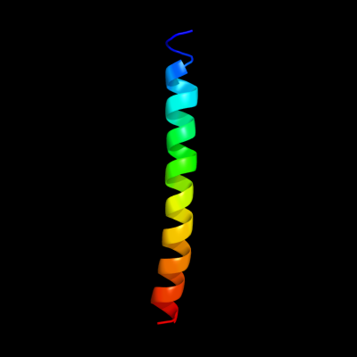
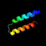
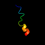
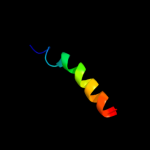
PDB header:blood clotting Chain: A: PDB Molecule:fibrin alpha-1 chain; PDBTitle: fibrin d-dimer, lamprey complexed with the peptide ligand: gly-his-2 arg-pro-amide
Confidence and coverage
Confidence:
62.8%
Coverage:
23%
34 residues ( 23% of your sequence) have been modelled with 62.8% confidence by the single highest scoring template.
You may wish to submit your sequence to Phyrealarm. This will automatically scan your sequence every week for new potential templates as they appear in the Phyre2 library.
Please note: You must be registered and logged in to use Phyrealarm.
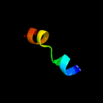
Region: 132 - 147 Aligned: 16 Modelled: 16 Confidence: 11.4% Identity: 13% PDB header:cell cycle protein Chain: B: PDB Molecule:iq2 motif from myo2p, a class v myosin; PDBTitle: crystal structure of mlc1p bound to iq2 of myo2p, a class v2 myosin
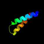
Region: 1 - 19 Aligned: 19 Modelled: 19 Confidence: 9.7% Identity: 16% Fold: Single transmembrane helix Superfamily: Subunit III of photosystem I reaction centre, PsaF Family: Subunit III of photosystem I reaction centre, PsaF
Region: 1 - 19 Aligned: 19 Modelled: 19 Confidence: 9.4% Identity: 11% PDB header:photosynthesis Chain: F: PDB Molecule:photosystem i reaction center subunit iii, PDBTitle: the structure of a plant photosystem i supercomplex at 3.42 angstrom resolution