| Secondary structure and disorder prediction | |
| | |
1 | . | . | . | . | . | . | . | . | 10 | . | . | . | . | . | . | . | . | . | 20 | . | . | . | . | . | . | . | . | . | 30 | . | . | . | . | . | . | . | . | . | 40 | . | . | . | . | . | . | . | . | . | 50 | . | . | . | . | . | . | . | . | . | 60 |
| Sequence | |
M | N | K | D | E | A | G | G | N | W | K | Q | F | K | G | K | V | K | E | Q | W | G | K | L | T | D | D | D | M | T | I | I | E | G | K | R | D | Q | L | V | G | K | I | Q | E | R | Y | G | Y | Q | K | D | Q | A | E | K | E | V | V | D |
| Secondary structure | |
|
|  | | | | | | | | | | | | | | | | | | | | | |
|
|
| | | | | |
|
|
| | | | | | | | | | | | |
| | | | | | | | | | | | |
| SS confidence | |
|
|
|
|
|
|
|
|
|
|
|
|
|
|
|
|
|
|
|
|
|
|
|
|
|
|
|
|
|
|
|
|
|
|
|
|
|
|
|
|
|
|
|
|
|
|
|
|
|
|
|
|
|
|
|
|
|
|
|
|
| Disorder | |
? | ? |
|
|
|
| ? |
|
|
|
|
|
|
|
|
|
|
|
|
|
|
|
|
|
|
|
|
|
|
|
|
| ? |
|
|
|
|
|
|
|
|
|
|
|
|
|
|
| ? |
|
|
|
|
|
|
|
|
|
|
|
| Disorder confidence | |
|
|
|
|
|
|
|
|
|
|
|
|
|
|
|
|
|
|
|
|
|
|
|
|
|
|
|
|
|
|
|
|
|
|
|
|
|
|
|
|
|
|
|
|
|
|
|
|
|
|
|
|
|
|
|
|
|
|
|
|
| |
| | |
. | . | . | . | . | . | . | . | . |
| Sequence | |
W | E | T | R | N | E | Y | R | W |
| Secondary structure | |
| | |
|
|
|
|
|
|
| SS confidence | |
|
|
|
|
|
|
|
|
|
| Disorder | |
|
| ? | ? | ? | ? | ? | ? | ? |
| Disorder confidence | |
|
|
|
|
|
|
|
|
|
| |
| Confidence Key |
| High(9) | |
|
|
|
|
|
|
|
|
|
Low (0) |
| ? | Disordered |
| Alpha helix |
  | Beta strand |
Hover over an aligned region to see model and summary info
Please note, only up to the top 20 hits are modelled to reduce computer load
|
| 1 |
|
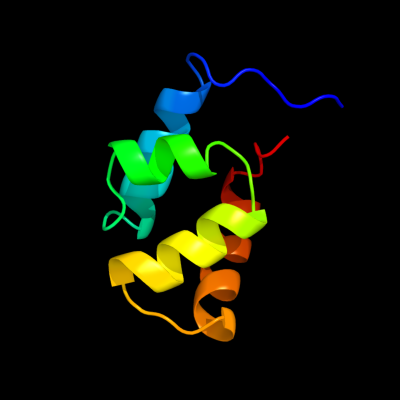
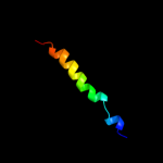
PDB 1ryk chain A
Region: 1 - 69
Aligned: 69
Modelled: 69
Confidence: 100.0%
Identity: 100%
Fold: SAM domain-like
Superfamily: Hypothetical protein YjbJ
Family: Hypothetical protein YjbJ
Phyre2
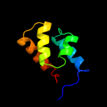
| 2 |
|
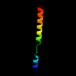
PDB 1yww chain A
Region: 1 - 64
Aligned: 64
Modelled: 64
Confidence: 99.9%
Identity: 55%
PDB header:structural genomics, unknown function
Chain: A: PDB Molecule:hypothetical protein pa4738;
PDBTitle: nmr structure of p. aeruginosa protein pa4738: northeast2 structural genomics consortium target pap2
Phyre2
| 3 |
|
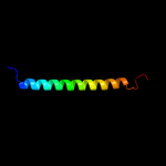
PDB 2l9q chain A
Region: 3 - 66
Aligned: 64
Modelled: 64
Confidence: 16.1%
Identity: 19%
PDB header:chaperone
Chain: A: PDB Molecule:12 kda heat shock protein;
PDBTitle: structural characterization of small heat shock protein (hsp12)
Phyre2
| 4 |
|
PDB 2js5 chain B
Region: 37 - 67
Aligned: 31
Modelled: 31
Confidence: 9.8%
Identity: 16%
PDB header:structural genomics, unknown function
Chain: B: PDB Molecule:uncharacterized protein;
PDBTitle: nmr structure of protein q60c73_metca. northeast structural2 genomics consortium target mcr1
Phyre2
| 5 |
|
PDB 2k88 chain A
Region: 29 - 66
Aligned: 38
Modelled: 38
Confidence: 8.4%
Identity: 21%
PDB header:hydrolase
Chain: A: PDB Molecule:vacuolar proton pump subunit g;
PDBTitle: association of subunit d (vma6p) and e (vma4p) with g2 (vma10p) and the nmr solution structure of subunit g (g1-3 59) of the saccharomyces cerevisiae v1vo atpase
Phyre2
| 6 |
|
PDB 3lf9 chain A
Region: 22 - 67
Aligned: 46
Modelled: 46
Confidence: 5.7%
Identity: 11%
PDB header:immune system
Chain: A: PDB Molecule:4e10_d0_1is1a_001_c (t161);
PDBTitle: crystal structure of hiv epitope-scaffold 4e10_d0_1is1a_001_c
Phyre2
|
| Detailed template information | |
Due to computational demand, binding site predictions are not run for batch jobs
If you want to predict binding sites, please manually submit your model of choice to 3DLigandSite
Phyre is for academic use only
| Please cite: Protein structure prediction on
the web: a case study using the Phyre server |
| Kelley LA and Sternberg MJE. Nature Protocols
4, 363 - 371 (2009) [pdf] [Import into BibTeX] |
| |
| If you use the binding site
predictions from 3DLigandSite, please also cite: |
| 3DLigandSite: predicting ligand-binding sites using similar structures. |
| Wass MN, Kelley LA and Sternberg
MJ Nucleic Acids Research 38, W469-73 (2010) [PubMed] |
| |
|
|
|
|