Phyre Home Retrieve Phyre Job Id
Emaill.a.kelley@imperial.ac.uk
DescriptionP0ADB1
DateThu Jan 5 11:20:30 GMT 2012
Unique Job IDc08444166a67d157

Summary 

Top model
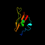
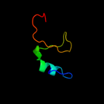
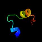
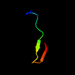
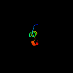
Image coloured by rainbow N → C terminus
Model (left) based on template c2pxgA_
Top template information
PDB header:membrane protein
Chain: A: PDB Molecule:outer membrane protein;
PDBTitle: nmr solution structure of omla
Confidence and coverage
Confidence: 96.9% Coverage: 69%
77 residues ( 69% of your sequence) have been modelled with 96.9% confidence by the single highest scoring template.
You may wish to submit your sequence to Phyrealarm. This will automatically scan your sequence every week for new potential templates as they appear in the Phyre2 library.
3D viewing
Interactive 3D view in Jmol

Sequence analysis 

Secondary structure and disorder prediction 

   1........10.........20.........30.........40.........50.........60
Sequence  MNKNMAGILSAAAVLTMLAGCTAYDRTKDQFVQPVVKDVKKGMSRAQVAQIAGKPSSEVS
Secondary structure 


SS confidence 


Disorder  ????


??
??


?


?


Disorder confidence 


   .........70.........80.........90.........100.........110..
Sequence  MIHARGTCQTYILGQRDGKAETYFVALDDTGHVINSGYQTCAEYDTDPQAAK
Secondary structure 


SS confidence 


Disorder 

?


????


?


????????
Disorder confidence 


Confidence Key
High(9)                    Low (0)
?Disordered
Alpha helix
Beta strand

Domain analysis 

Hover over an aligned region to see model and summary info

Please note, only up to the top 20 hits are modelled to reduce computer load

RankAligned region

PDB 2pxg chain A

3D model

Region: 18 - 97
Aligned: 77
Modelled: 80
Confidence: 96.9%
Identity: 23%
PDB header:membrane protein
Chain: A: PDB Molecule:outer membrane protein;
PDBTitle: nmr solution structure of omla

Phyre2

PDB 2kxx chain A

3D model

Region: 36 - 95
Aligned: 58
Modelled: 60
Confidence: 96.5%
Identity: 22%
PDB header:protein binding
Chain: A: PDB Molecule:small protein a;
PDBTitle: nmr structure of escherichia coli bame, a lipoprotein component of the2 beta-barrel assembly machinery complex

Phyre2

PDB 3gmx chain B

3D model

Region: 42 - 96
Aligned: 50
Modelled: 55
Confidence: 79.0%
Identity: 24%
PDB header:protein binding
Chain: B: PDB Molecule:blp;
PDBTitle: crystal structure of beta-lactamse inhibitory protein-like2 protein (blp) at 1.05 angstrom resolution

Phyre2

PDB 3d4e chain A

3D model

Region: 38 - 87
Aligned: 50
Modelled: 50
Confidence: 74.7%
Identity: 14%
PDB header:hydrolase
Chain: A: PDB Molecule:putative beta-lactamase inhibitor protein;
PDBTitle: crystal structure of putative beta-lactamase inhibitor protein2 (np_721579.1) from streptococcus mutans at 1.40 a resolution

Phyre2

PDB 2g2u chain B domain 1

3D model

Region: 37 - 78
Aligned: 41
Modelled: 42
Confidence: 67.2%
Identity: 24%
Fold: BLIP-like
Superfamily: beta-lactamase-inhibitor protein, BLIP
Family: beta-lactamase-inhibitor protein, BLIP

Phyre2

PDB 3gmv chain X

3D model

Region: 42 - 53
Aligned: 12
Modelled: 12
Confidence: 64.4%
Identity: 42%
PDB header:protein binding
Chain: X: PDB Molecule:beta-lactamase inhibitory protein blip-i;
PDBTitle: crystal structure of beta-lactamse inhibitory protein-i2 (blip-i) in apo form

Phyre2

PDB 2w7n chain A

3D model

Region: 36 - 51
Aligned: 16
Modelled: 16
Confidence: 19.8%
Identity: 25%
PDB header:transcription/dna
Chain: A: PDB Molecule:trfb transcriptional repressor protein;
PDBTitle: crystal structure of kora bound to operator dna: insight2 into repressor cooperation in rp4 gene regulation

Phyre2

PDB 2ppx chain A domain 1

3D model

Region: 34 - 57
Aligned: 24
Modelled: 24
Confidence: 17.6%
Identity: 13%
Fold: lambda repressor-like DNA-binding domains
Superfamily: lambda repressor-like DNA-binding domains
Family: SinR domain-like

Phyre2

PDB 2ppx chain A

3D model

Region: 34 - 57
Aligned: 24
Modelled: 24
Confidence: 17.6%
Identity: 13%
PDB header:structural genomics, unknown function
Chain: A: PDB Molecule:uncharacterized protein atu1735;
PDBTitle: crystal structure of a hth xre-family like protein from agrobacterium2 tumefaciens

Phyre2

PDB 2fhd chain A

3D model

Region: 65 - 94
Aligned: 29
Modelled: 27
Confidence: 14.6%
Identity: 17%
PDB header:cell cycle
Chain: A: PDB Molecule:dna repair protein rhp9/crb2;
PDBTitle: crystal structure of crb2 tandem tudor domains

Phyre2

PDB 3d0w chain D

3D model

Region: 22 - 52
Aligned: 31
Modelled: 31
Confidence: 11.9%
Identity: 16%
PDB header:structural genomics, unknown function
Chain: D: PDB Molecule:yflh protein;
PDBTitle: crystal structure of yflh protein from bacillus subtilis.2 northeast structural genomics consortium target sr326

Phyre2

PDB 2a7y chain A

3D model

Region: 77 - 94
Aligned: 17
Modelled: 18
Confidence: 11.4%
Identity: 35%
PDB header:structural genomics, unknown function
Chain: A: PDB Molecule:hypothetical protein rv2302/mt2359;
PDBTitle: solution structure of the conserved hypothetical protein2 rv2302 from the bacterium mycobacterium tuberculosis

Phyre2

PDB 2a7y chain A domain 1

3D model

Region: 77 - 94
Aligned: 17
Modelled: 18
Confidence: 11.4%
Identity: 35%
Fold: SH3-like barrel
Superfamily: Cell growth inhibitor/plasmid maintenance toxic component
Family: Rv2302-like

Phyre2

PDB 1bw6 chain A

3D model

Region: 35 - 58
Aligned: 24
Modelled: 24
Confidence: 10.1%
Identity: 17%
Fold: DNA/RNA-binding 3-helical bundle
Superfamily: Homeodomain-like
Family: Centromere-binding

Phyre2

PDB 2kq5 chain A

3D model

Region: 34 - 57
Aligned: 23
Modelled: 24
Confidence: 9.7%
Identity: 30%
PDB header:unknown function
Chain: A: PDB Molecule:avirulence protein;
PDBTitle: solution nmr structure of a section of the repeat domain of the type2 iii effector protein ptha

Phyre2

PDB 2xzm chain O

3D model

Region: 21 - 48
Aligned: 28
Modelled: 28
Confidence: 9.1%
Identity: 29%
PDB header:ribosome
Chain: O: PDB Molecule:rps13e;
PDBTitle: crystal structure of the eukaryotic 40s ribosomal2 subunit in complex with initiation factor 1. this file3 contains the 40s subunit and initiation factor for4 molecule 1

Phyre2

PDB 3f6w chain E

3D model

Region: 40 - 59
Aligned: 20
Modelled: 20
Confidence: 8.7%
Identity: 30%
PDB header:dna binding protein
Chain: E: PDB Molecule:xre-family like protein;
PDBTitle: xre-family like protein from pseudomonas syringae pv. tomato str.2 dc3000

Phyre2

PDB 1tfr chain A domain 1

3D model

Region: 69 - 82
Aligned: 14
Modelled: 14
Confidence: 8.7%
Identity: 7%
Fold: SAM domain-like
Superfamily: 5' to 3' exonuclease, C-terminal subdomain
Family: 5' to 3' exonuclease, C-terminal subdomain

Phyre2

PDB 3eip chain A

3D model

Region: 69 - 88
Aligned: 20
Modelled: 20
Confidence: 8.4%
Identity: 40%
Fold: FKBP-like
Superfamily: Colicin E3 immunity protein
Family: Colicin E3 immunity protein

Phyre2

PDB 3o4a chain C

3D model

Region: 79 - 96
Aligned: 18
Modelled: 18
Confidence: 7.9%
Identity: 11%
PDB header:de novo protein
Chain: C: PDB Molecule:de novo designed beta-trefoil architecture with symmetric
PDBTitle: crystal structure of symfoil-2: de novo designed beta-trefoil2 architecture with symmetric primary structure

Phyre2
1

c2pxgA_
2

c2kxxA_
3

c3gmxB_
4

c3d4eA_
5

d2g2ub1
6

c3gmvX_
7

c2w7nA_
8

d2ppxa1
9

c2ppxA_
10

c2fhdA_
11

c3d0wD_
12

c2a7yA_
13

d2a7ya1
14

d1bw6a_
15

c2kq5A_
16

c2xzmO_
17

c3f6wE_
18

d1tfra1
19

d3eipa_
20

c3o4aC_
21


22


23


24


25


26


27


28


29


30


31


32


33


34


35


Detailed template information 

#
Template Alignment Coverage3D Model Confidence
% i.d. Template Information
1c2pxgA_


96.9 23 PDB header:membrane protein
Chain: A: PDB Molecule:outer membrane protein;
PDBTitle: nmr solution structure of omla
2c2kxxA_


96.5 22 PDB header:protein binding
Chain: A: PDB Molecule:small protein a;
PDBTitle: nmr structure of escherichia coli bame, a lipoprotein component of the2 beta-barrel assembly machinery complex
3c3gmxB_


79.0 24 PDB header:protein binding
Chain: B: PDB Molecule:blp;
PDBTitle: crystal structure of beta-lactamse inhibitory protein-like2 protein (blp) at 1.05 angstrom resolution
4c3d4eA_


74.7 14 PDB header:hydrolase
Chain: A: PDB Molecule:putative beta-lactamase inhibitor protein;
PDBTitle: crystal structure of putative beta-lactamase inhibitor protein2 (np_721579.1) from streptococcus mutans at 1.40 a resolution
5d2g2ub1


67.2 24 Fold:BLIP-like
Superfamily:beta-lactamase-inhibitor protein, BLIP
Family:beta-lactamase-inhibitor protein, BLIP
6c3gmvX_


64.4 42 PDB header:protein binding
Chain: X: PDB Molecule:beta-lactamase inhibitory protein blip-i;
PDBTitle: crystal structure of beta-lactamse inhibitory protein-i2 (blip-i) in apo form
7c2w7nA_


19.8 25 PDB header:transcription/dna
Chain: A: PDB Molecule:trfb transcriptional repressor protein;
PDBTitle: crystal structure of kora bound to operator dna: insight2 into repressor cooperation in rp4 gene regulation
8d2ppxa1


17.6 13 Fold:lambda repressor-like DNA-binding domains
Superfamily:lambda repressor-like DNA-binding domains
Family:SinR domain-like
9c2ppxA_


17.6 13 PDB header:structural genomics, unknown function
Chain: A: PDB Molecule:uncharacterized protein atu1735;
PDBTitle: crystal structure of a hth xre-family like protein from agrobacterium2 tumefaciens
10c2fhdA_


14.6 17 PDB header:cell cycle
Chain: A: PDB Molecule:dna repair protein rhp9/crb2;
PDBTitle: crystal structure of crb2 tandem tudor domains
11c3d0wD_


11.9 16 PDB header:structural genomics, unknown function
Chain: D: PDB Molecule:yflh protein;
PDBTitle: crystal structure of yflh protein from bacillus subtilis.2 northeast structural genomics consortium target sr326
12c2a7yA_


11.4 35 PDB header:structural genomics, unknown function
Chain: A: PDB Molecule:hypothetical protein rv2302/mt2359;
PDBTitle: solution structure of the conserved hypothetical protein2 rv2302 from the bacterium mycobacterium tuberculosis
13d2a7ya1


11.4 35 Fold:SH3-like barrel
Superfamily:Cell growth inhibitor/plasmid maintenance toxic component
Family:Rv2302-like
14d1bw6a_


10.1 17 Fold:DNA/RNA-binding 3-helical bundle
Superfamily:Homeodomain-like
Family:Centromere-binding
15c2kq5A_


9.7 30 PDB header:unknown function
Chain: A: PDB Molecule:avirulence protein;
PDBTitle: solution nmr structure of a section of the repeat domain of the type2 iii effector protein ptha
16c2xzmO_


9.1 29 PDB header:ribosome
Chain: O: PDB Molecule:rps13e;
PDBTitle: crystal structure of the eukaryotic 40s ribosomal2 subunit in complex with initiation factor 1. this file3 contains the 40s subunit and initiation factor for4 molecule 1
17c3f6wE_


8.7 30 PDB header:dna binding protein
Chain: E: PDB Molecule:xre-family like protein;
PDBTitle: xre-family like protein from pseudomonas syringae pv. tomato str.2 dc3000
18d1tfra1


8.7 7 Fold:SAM domain-like
Superfamily:5' to 3' exonuclease, C-terminal subdomain
Family:5' to 3' exonuclease, C-terminal subdomain
19d3eipa_


8.4 40 Fold:FKBP-like
Superfamily:Colicin E3 immunity protein
Family:Colicin E3 immunity protein
20c3o4aC_


7.9 11 PDB header:de novo protein
Chain: C: PDB Molecule:de novo designed beta-trefoil architecture with symmetric
PDBTitle: crystal structure of symfoil-2: de novo designed beta-trefoil2 architecture with symmetric primary structure
21d1ihka_


not modelled 7.7 31 Fold:beta-Trefoil
Superfamily:Cytokine
Family:Fibroblast growth factors (FGF)
22c3mtjA_


not modelled 7.2 28 PDB header:oxidoreductase
Chain: A: PDB Molecule:homoserine dehydrogenase;
PDBTitle: the crystal structure of a homoserine dehydrogenase from thiobacillus2 denitrificans to 2.15a
23d2oq1a2


not modelled 7.2 18 Fold:SH2-like
Superfamily:SH2 domain
Family:SH2 domain
24d2a6ca1


not modelled 7.0 19 Fold:lambda repressor-like DNA-binding domains
Superfamily:lambda repressor-like DNA-binding domains
Family:NE1354
25c3f1rA_


not modelled 7.0 38 PDB header:hormone
Chain: A: PDB Molecule:fibroblast growth factor 20;
PDBTitle: crystal structure of fgf20 dimer
26d1ijwc_


not modelled 7.0 50 Fold:DNA/RNA-binding 3-helical bundle
Superfamily:Homeodomain-like
Family:Recombinase DNA-binding domain
27d1f35a_


not modelled 6.9 40 Fold:Olfactory marker protein
Superfamily:Olfactory marker protein
Family:Olfactory marker protein
28c3bs3A_


not modelled 6.4 22 PDB header:dna binding protein
Chain: A: PDB Molecule:putative dna-binding protein;
PDBTitle: crystal structure of a putative dna-binding protein from bacteroides2 fragilis
29d2hjqa1


not modelled 6.3 38 Fold:LEM/SAP HeH motif
Superfamily:Rho N-terminal domain-like
Family:YqbF C-terminal domain-like
30d2axwa1


not modelled 6.0 9 Fold:Common fold of diphtheria toxin/transcription factors/cytochrome f
Superfamily:Bacterial adhesins
Family:Pilus subunits
31d1pxza_


not modelled 5.6 13 Fold:Single-stranded right-handed beta-helix
Superfamily:Pectin lyase-like
Family:Pectate lyase-like
32c2iurD_


not modelled 5.6 41 PDB header:oxidoreductase
Chain: D: PDB Molecule:aromatic amine dehydrogenase beta subunit;
PDBTitle: crystal structure of n-quinol form of aromatic amine2 dehydrogenase (aadh) from alcaligenes faecalis, form a3 cocrystal
33c2p04B_


not modelled 5.3 13 PDB header:transferase
Chain: B: PDB Molecule:signal transduction histidine kinase;
PDBTitle: 2.1 ang structure of the dimerized pas domain of signal transduction2 histidine kinase from nostoc punctiforme pcc 73102 with homology to3 the h-noxa/h-noba domain of the soluble guanylyl cyclase
34c1w78A_


not modelled 5.2 15 PDB header:synthase
Chain: A: PDB Molecule:folc bifunctional protein;
PDBTitle: e.coli folc in complex with dhpp and adp
35c2o1uA_


not modelled 5.2 44 PDB header:chaperone
Chain: A: PDB Molecule:endoplasmin;
PDBTitle: structure of full length grp94 with amp-pnp bound

Binding site prediction 

Due to computational demand, binding site predictions are not run for batch jobs

If you want to predict binding sites, please manually submit your model of choice to 3DLigandSite


Phyre is for academic use only

Please cite: Protein structure prediction on the web: a case study using the Phyre server
Kelley LA and Sternberg MJE. Nature Protocols 4, 363 - 371 (2009) [pdf] [Import into BibTeX]
 
If you use the binding site predictions from 3DLigandSite, please also cite:
3DLigandSite: predicting ligand-binding sites using similar structures.
Wass MN, Kelley LA and Sternberg MJ Nucleic Acids Research 38, W469-73 (2010) [PubMed]
 
© Structural Bioinformatics Group
Imperial College London
Lawrence Kelley, Benjamin Jefferys
Disclaimer
Terms and Conditions
Component software
Template detection: HHpred 1.51
Secondary structure prediction: Psi-pred 2.5
Disorder prediction: Disopred 2.4
Transmembrane prediction: Memsat_SVM
Multi-template modelling and ab initio: Poing 1.0