Phyre Home Retrieve Phyre Job Id
Emaill.a.kelley@imperial.ac.uk
DescriptionP0A7Y4
DateThu Jan 5 11:06:28 GMT 2012
Unique Job IDbecbb231fdaf04d1

Summary 

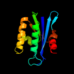
Top model
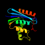
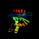
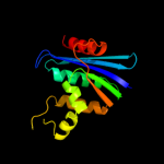
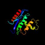
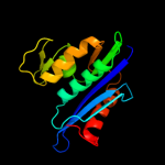
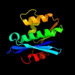
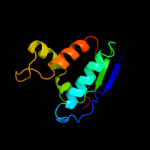
Image coloured by rainbow N → C terminus
Model (left) based on template d1jl1a_
Top template information
Fold:Ribonuclease H-like motif
Superfamily:Ribonuclease H-like
Family:Ribonuclease H
Confidence and coverage
Confidence:100.0% Coverage: 98%
152 residues ( 98% of your sequence) have been modelled with 100.0% confidence by the single highest scoring template.
3D viewing
Interactive 3D view in Jmol

Sequence analysis 

Secondary structure and disorder prediction 

   1........10.........20.........30.........40.........50.........60
Sequence  MLKQVEIFTDGSCLGNPGPGGYGAILRYRGREKTFSAGYTRTTNNRMELMAAIVALEALK
Secondary structure 


SS confidence 


Disorder  ???


?


?

???


Disorder confidence 


   .........70.........80.........90.........100.........110.........120
Sequence  EHCEVILSTDSQYVRQGITQWIHNWKKRGWKTADKKPVKNVDLWQRLDAALGQHQIKWEW
Secondary structure 


SS confidence 


Disorder 


??
??????

?


Disorder confidence 


   .........130.........140.........150.....
Sequence  VKGHAGHPENERCDELARAAAMNPTLEDTGYQVEV
Secondary structure 


SS confidence 


Disorder 


???????????????
Disorder confidence 


Confidence Key
High(9)                    Low (0)
?Disordered
Alpha helix
Beta strand

Domain analysis 

Hover over an aligned region to see model and summary info

Please note, only up to the top 20 hits are modelled to reduce computer load

RankAligned region

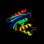
PDB 1jl1 chain A

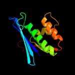
3D model

Region: 3 - 154
Aligned: 152
Modelled: 152
Confidence: 100.0%
Identity: 97%
Fold: Ribonuclease H-like motif
Superfamily: Ribonuclease H-like
Family: Ribonuclease H

Phyre2

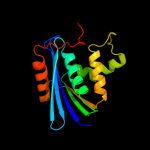
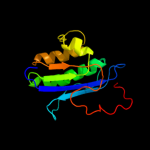
PDB 1jl2 chain A

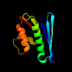
3D model

Region: 3 - 151
Aligned: 149
Modelled: 149
Confidence: 100.0%
Identity: 74%
Fold: Ribonuclease H-like motif
Superfamily: Ribonuclease H-like
Family: Ribonuclease H

Phyre2

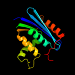
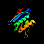
PDB 2e4l chain A

3D model

Region: 2 - 154
Aligned: 153
Modelled: 153
Confidence: 100.0%
Identity: 70%
PDB header:hydrolase
Chain: A: PDB Molecule:ribonuclease hi;
PDBTitle: thermodynamic and structural analysis of thermolabile rnase hi from2 shewanella oneidensis mr-1

Phyre2

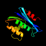
PDB 1wsi chain B

3D model

Region: 2 - 153
Aligned: 149
Modelled: 152
Confidence: 100.0%
Identity: 99%
Fold: Ribonuclease H-like motif
Superfamily: Ribonuclease H-like
Family: Ribonuclease H

Phyre2

PDB 1ril chain A

3D model

Region: 2 - 145
Aligned: 144
Modelled: 144
Confidence: 100.0%
Identity: 56%
Fold: Ribonuclease H-like motif
Superfamily: Ribonuclease H-like
Family: Ribonuclease H

Phyre2

PDB 3h08 chain B

3D model

Region: 3 - 142
Aligned: 139
Modelled: 140
Confidence: 100.0%
Identity: 45%
PDB header:hydrolase
Chain: B: PDB Molecule:rnh (ribonuclease h);
PDBTitle: crystal structure of the ribonuclease h1 from chlorobium2 tepidum

Phyre2

PDB 2qkk chain I

3D model

Region: 1 - 144
Aligned: 144
Modelled: 144
Confidence: 100.0%
Identity: 35%
PDB header:hydrolase/dna/rna
Chain: I: PDB Molecule:ribonuclease h1;
PDBTitle: human rnase h catalytic domain mutant d210n in complex with2 14-mer rna/dna hybrid

Phyre2

PDB 2hb5 chain A

3D model

Region: 3 - 149
Aligned: 134
Modelled: 147
Confidence: 100.0%
Identity: 28%
PDB header:hydrolase
Chain: A: PDB Molecule:reverse transcriptase/ribonuclease h;
PDBTitle: crystal structure of the moloney murine leukemia virus2 rnase h domain

Phyre2

PDB 1eet chain A domain 1

3D model

Region: 5 - 142
Aligned: 120
Modelled: 125
Confidence: 99.9%
Identity: 27%
Fold: Ribonuclease H-like motif
Superfamily: Ribonuclease H-like
Family: Ribonuclease H

Phyre2

PDB 1mu2 chain A domain 1

3D model

Region: 4 - 140
Aligned: 119
Modelled: 124
Confidence: 99.9%
Identity: 26%
Fold: Ribonuclease H-like motif
Superfamily: Ribonuclease H-like
Family: Ribonuclease H

Phyre2

PDB 1jla chain A domain 1

3D model

Region: 1 - 137
Aligned: 119
Modelled: 124
Confidence: 99.9%
Identity: 26%
Fold: Ribonuclease H-like motif
Superfamily: Ribonuclease H-like
Family: Ribonuclease H

Phyre2

PDB 1zbf chain A domain 1

3D model

Region: 7 - 134
Aligned: 124
Modelled: 128
Confidence: 99.9%
Identity: 21%
Fold: Ribonuclease H-like motif
Superfamily: Ribonuclease H-like
Family: Ribonuclease H

Phyre2

PDB 2kq2 chain A

3D model

Region: 4 - 147
Aligned: 129
Modelled: 129
Confidence: 99.9%
Identity: 22%
PDB header:hydrolase
Chain: A: PDB Molecule:ribonuclease h-related protein;
PDBTitle: solution nmr structure of the apo form of a ribonuclease h2 domain of protein dsy1790 from desulfitobacterium3 hafniense, northeast structural genomics target dhr1a

Phyre2

PDB 2rpi chain A

3D model

Region: 3 - 122
Aligned: 99
Modelled: 99
Confidence: 99.9%
Identity: 63%
PDB header:hydrolase
Chain: A: PDB Molecule:ribonuclease h;
PDBTitle: the nmr structure of the submillisecond folding2 intermediate of the thermus thermophilus ribonuclease h

Phyre2

PDB 1o1w chain A

3D model

Region: 1 - 143
Aligned: 125
Modelled: 130
Confidence: 99.9%
Identity: 26%
Fold: Ribonuclease H-like motif
Superfamily: Ribonuclease H-like
Family: Ribonuclease H

Phyre2

PDB 3hst chain D

3D model

Region: 4 - 142
Aligned: 125
Modelled: 139
Confidence: 99.9%
Identity: 31%
PDB header:hydrolase
Chain: D: PDB Molecule:protein rv2228c/mt2287;
PDBTitle: n-terminal rnase h domain of rv2228c from mycobacterium tuberculosis2 as a fusion protein with maltose binding protein

Phyre2

PDB 1mu2 chain A

3D model

Region: 5 - 140
Aligned: 118
Modelled: 121
Confidence: 99.9%
Identity: 25%
PDB header:transferase
Chain: A: PDB Molecule:hiv-2 rt;
PDBTitle: crystal structure of hiv-2 reverse transcriptase

Phyre2

PDB 2ehg chain A

3D model

Region: 5 - 142
Aligned: 123
Modelled: 126
Confidence: 99.8%
Identity: 24%
PDB header:hydrolase
Chain: A: PDB Molecule:ribonuclease hi;
PDBTitle: crystal structure of hyperthermophilic archaeal rnase hi

Phyre2

PDB 1s9g chain A domain 1

3D model

Region: 5 - 127
Aligned: 105
Modelled: 110
Confidence: 99.8%
Identity: 26%
Fold: Ribonuclease H-like motif
Superfamily: Ribonuclease H-like
Family: Ribonuclease H

Phyre2

PDB 1s1t chain A domain 1

3D model

Region: 5 - 124
Aligned: 102
Modelled: 107
Confidence: 99.8%
Identity: 25%
Fold: Ribonuclease H-like motif
Superfamily: Ribonuclease H-like
Family: Ribonuclease H

Phyre2
1

d1jl1a_
2

d1jl2a_
3

c2e4lA_
4

d1wsib_
5

d1rila_
6

c3h08B_
7

c2qkkI_
8

c2hb5A_
9

d1eeta1
10

d1mu2a1
11

d1jlaa1
12

d1zbfa1
13

c2kq2A_
14

c2rpiA_
15

d1o1wa_
16

c3hstD_
17

c1mu2A_
18

c2ehgA_
19

d1s9ga1
20

d1s1ta1
21


22


23


24


25


26


27


28


29


30


31


32


33


34


Detailed template information 

#
Template Alignment Coverage3D Model Confidence
% i.d. Template Information
1d1jl1a_


100.0 97 Fold:Ribonuclease H-like motif
Superfamily:Ribonuclease H-like
Family:Ribonuclease H
2d1jl2a_


100.0 74 Fold:Ribonuclease H-like motif
Superfamily:Ribonuclease H-like
Family:Ribonuclease H
3c2e4lA_


100.0 70 PDB header:hydrolase
Chain: A: PDB Molecule:ribonuclease hi;
PDBTitle: thermodynamic and structural analysis of thermolabile rnase hi from2 shewanella oneidensis mr-1
4d1wsib_


100.0 99 Fold:Ribonuclease H-like motif
Superfamily:Ribonuclease H-like
Family:Ribonuclease H
5d1rila_


100.0 56 Fold:Ribonuclease H-like motif
Superfamily:Ribonuclease H-like
Family:Ribonuclease H
6c3h08B_


100.0 45 PDB header:hydrolase
Chain: B: PDB Molecule:rnh (ribonuclease h);
PDBTitle: crystal structure of the ribonuclease h1 from chlorobium2 tepidum
7c2qkkI_


100.0 35 PDB header:hydrolase/dna/rna
Chain: I: PDB Molecule:ribonuclease h1;
PDBTitle: human rnase h catalytic domain mutant d210n in complex with2 14-mer rna/dna hybrid
8c2hb5A_


100.0 28 PDB header:hydrolase
Chain: A: PDB Molecule:reverse transcriptase/ribonuclease h;
PDBTitle: crystal structure of the moloney murine leukemia virus2 rnase h domain
9d1eeta1


99.9 27 Fold:Ribonuclease H-like motif
Superfamily:Ribonuclease H-like
Family:Ribonuclease H
10d1mu2a1


99.9 26 Fold:Ribonuclease H-like motif
Superfamily:Ribonuclease H-like
Family:Ribonuclease H
11d1jlaa1


99.9 26 Fold:Ribonuclease H-like motif
Superfamily:Ribonuclease H-like
Family:Ribonuclease H
12d1zbfa1


99.9 21 Fold:Ribonuclease H-like motif
Superfamily:Ribonuclease H-like
Family:Ribonuclease H
13c2kq2A_


99.9 22 PDB header:hydrolase
Chain: A: PDB Molecule:ribonuclease h-related protein;
PDBTitle: solution nmr structure of the apo form of a ribonuclease h2 domain of protein dsy1790 from desulfitobacterium3 hafniense, northeast structural genomics target dhr1a
14c2rpiA_


99.9 63 PDB header:hydrolase
Chain: A: PDB Molecule:ribonuclease h;
PDBTitle: the nmr structure of the submillisecond folding2 intermediate of the thermus thermophilus ribonuclease h
15d1o1wa_


99.9 26 Fold:Ribonuclease H-like motif
Superfamily:Ribonuclease H-like
Family:Ribonuclease H
16c3hstD_


99.9 31 PDB header:hydrolase
Chain: D: PDB Molecule:protein rv2228c/mt2287;
PDBTitle: n-terminal rnase h domain of rv2228c from mycobacterium tuberculosis2 as a fusion protein with maltose binding protein
17c1mu2A_


99.9 25 PDB header:transferase
Chain: A: PDB Molecule:hiv-2 rt;
PDBTitle: crystal structure of hiv-2 reverse transcriptase
18c2ehgA_


99.8 24 PDB header:hydrolase
Chain: A: PDB Molecule:ribonuclease hi;
PDBTitle: crystal structure of hyperthermophilic archaeal rnase hi
19d1s9ga1


99.8 26 Fold:Ribonuclease H-like motif
Superfamily:Ribonuclease H-like
Family:Ribonuclease H
20d1s1ta1


99.8 25 Fold:Ribonuclease H-like motif
Superfamily:Ribonuclease H-like
Family:Ribonuclease H
21c1rthA_


not modelled 99.6 26 PDB header:nucleotidyltransferase
Chain: A: PDB Molecule:hiv-1 reverse transcriptase;
PDBTitle: high resolution structures of hiv-1 rt from four rt-2 inhibitor complexes
22c2opqA_


not modelled 99.2 27 PDB header:transferase
Chain: A: PDB Molecule:reverse transcriptase/ribonuclease h;
PDBTitle: crystal structure of l100i mutant hiv-1 reverse2 transcriptase in complex with gw420867x.
23d1rw3a_


not modelled 56.1 8 Fold:DNA/RNA polymerases
Superfamily:DNA/RNA polymerases
Family:Reverse transcriptase
24c2hxvA_


not modelled 22.7 17 PDB header:biosynthetic protein
Chain: A: PDB Molecule:diaminohydroxyphosphoribosylaminopyrimidine deaminase/ 5-
PDBTitle: crystal structure of a diaminohydroxyphosphoribosylaminopyrimidine2 deaminase/ 5-amino-6-(5-phosphoribosylamino)uracil reductase (tm1828)3 from thermotoga maritima at 1.80 a resolution
25d2b3za2


not modelled 13.1 16 Fold:Cytidine deaminase-like
Superfamily:Cytidine deaminase-like
Family:Deoxycytidylate deaminase-like
26c3hl4B_


not modelled 11.5 23 PDB header:transferase
Chain: B: PDB Molecule:choline-phosphate cytidylyltransferase a;
PDBTitle: crystal structure of a mammalian ctp:phosphocholine2 cytidylyltransferase with cdp-choline
27d1ynja1


not modelled 9.4 63 Fold:DCoH-like
Superfamily:RBP11-like subunits of RNA polymerase
Family:RNA polymerase alpha subunit dimerisation domain
28d1nuia2


not modelled 9.4 38 Fold:Rubredoxin-like
Superfamily:Zinc beta-ribbon
Family:DNA primase zinc finger
29d2g84a1


not modelled 9.2 19 Fold:Cytidine deaminase-like
Superfamily:Cytidine deaminase-like
Family:Deoxycytidylate deaminase-like
30d1mr4a_


not modelled 8.2 63 Fold:Knottins (small inhibitors, toxins, lectins)
Superfamily:Scorpion toxin-like
Family:Plant defensins
31d1cw0a_


not modelled 8.1 36 Fold:Restriction endonuclease-like
Superfamily:Restriction endonuclease-like
Family:Very short patch repair (VSR) endonuclease
32d1vsra_


not modelled 7.8 36 Fold:Restriction endonuclease-like
Superfamily:Restriction endonuclease-like
Family:Very short patch repair (VSR) endonuclease
33c2d5nB_


not modelled 7.3 22 PDB header:hydrolase, oxidoreductase
Chain: B: PDB Molecule:riboflavin biosynthesis protein ribd;
PDBTitle: crystal structure of a bifunctional deaminase and reductase2 involved in riboflavin biosynthesis
34d1smya1


not modelled 7.3 63 Fold:DCoH-like
Superfamily:RBP11-like subunits of RNA polymerase
Family:RNA polymerase alpha subunit dimerisation domain

Binding site prediction 

Due to computational demand, binding site predictions are not run for batch jobs

If you want to predict binding sites, please manually submit your model of choice to 3DLigandSite


Phyre is for academic use only

Please cite: Protein structure prediction on the web: a case study using the Phyre server
Kelley LA and Sternberg MJE. Nature Protocols 4, 363 - 371 (2009) [pdf] [Import into BibTeX]
 
If you use the binding site predictions from 3DLigandSite, please also cite:
3DLigandSite: predicting ligand-binding sites using similar structures.
Wass MN, Kelley LA and Sternberg MJ Nucleic Acids Research 38, W469-73 (2010) [PubMed]
 
© Structural Bioinformatics Group
Imperial College London
Lawrence Kelley, Benjamin Jefferys
Disclaimer
Terms and Conditions
Component software
Template detection: HHpred 1.51
Secondary structure prediction: Psi-pred 2.5
Disorder prediction: Disopred 2.4
Transmembrane prediction: Memsat_SVM
Multi-template modelling and ab initio: Poing 1.0