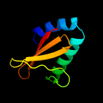
| 1 | d2a6sa1
|
|
 |
100.0 |
100 |
Fold:RelE-like
Superfamily:RelE-like
Family:YoeB/Txe-like |
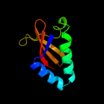
| 2 | c3oeiH_
|
|
 |
100.0 |
46 |
PDB header:toxin, protein binding
Chain: H: PDB Molecule:relk (toxin rv3358);
PDBTitle: crystal structure of mycobacterium tuberculosis reljk (rv3357-rv3358-2 relbe3)
|
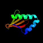
| 3 | d1z8ma1
|
|
 |
99.8 |
19 |
Fold:RelE-like
Superfamily:RelE-like
Family:RelE-like |
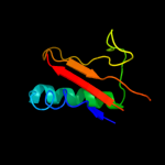
| 4 | c2otrA_
|
|
 |
99.8 |
18 |
PDB header:structural genomics, unknown function
Chain: A: PDB Molecule:hypothetical protein hp0892;
PDBTitle: solution structure of conserved hypothetical protein hp0892 from2 helicobacter pylori
|
| 5 | c2kheA_
|
|
 |
99.4 |
18 |
PDB header:hydrolase
Chain: A: PDB Molecule:toxin-like protein;
PDBTitle: solution structure of the bacterial toxin rele from thermus2 thermophilus hb8
|
| 6 | c3bpqD_
|
|
 |
99.3 |
13 |
PDB header:toxin
Chain: D: PDB Molecule:toxin rele3;
PDBTitle: crystal structure of relb-rele antitoxin-toxin complex from2 methanococcus jannaschii
|
| 7 | c3g5oC_
|
|
 |
99.3 |
25 |
PDB header:toxin/antitoxin
Chain: C: PDB Molecule:uncharacterized protein rv2866;
PDBTitle: the crystal structure of the toxin-antitoxin complex relbe2 (rv2865-2 2866) from mycobacterium tuberculosis
|
| 8 | d1wmia1
|
|
 |
99.3 |
14 |
Fold:RelE-like
Superfamily:RelE-like
Family:RelE-like |
| 9 | c3kixy_
|
|
 |
98.6 |
15 |
PDB header:ribosome
Chain: Y: PDB Molecule:
PDBTitle: structure of rele nuclease bound to the 70s ribosome2 (postcleavage state; part 3 of 4)
|
| 10 | c3kxeB_
|
|
 |
97.5 |
16 |
PDB header:protein binding
Chain: B: PDB Molecule:toxin protein pare-1;
PDBTitle: a conserved mode of protein recognition and binding in a2 pard-pare toxin-antitoxin complex
|
| 11 | d2p2ea1
|
|
 |
32.3 |
7 |
Fold:HesB-like domain
Superfamily:HesB-like domain
Family:HesB-like domain |
| 12 | c2qgoA_
|
|
 |
31.9 |
7 |
PDB header:biosynthetic protein
Chain: A: PDB Molecule:putative fe-s biosynthesis protein;
PDBTitle: crystal structure of a putative fe-s biosynthesis protein from2 lactobacillus acidophilus
|
| 13 | d3bb9a1
|
|
 |
24.1 |
7 |
Fold:Cystatin-like
Superfamily:NTF2-like
Family:SO0125-like |
| 14 | d2qn6a2
|
|
 |
22.0 |
36 |
Fold:Elongation factor/aminomethyltransferase common domain
Superfamily:EF-Tu/eEF-1alpha/eIF2-gamma C-terminal domain
Family:EF-Tu/eEF-1alpha/eIF2-gamma C-terminal domain |
| 15 | d1s0ua2
|
|
 |
21.9 |
45 |
Fold:Elongation factor/aminomethyltransferase common domain
Superfamily:EF-Tu/eEF-1alpha/eIF2-gamma C-terminal domain
Family:EF-Tu/eEF-1alpha/eIF2-gamma C-terminal domain |
| 16 | c2l09A_
|
|
 |
20.7 |
27 |
PDB header:oxidoreductase
Chain: A: PDB Molecule:asr4154 protein;
PDBTitle: solution nmr structure of protein asr4154 from nostoc sp. pcc71202 northeast structural genomics consortium target id nsr143
|
| 17 | d1kk1a2
|
|
 |
20.5 |
45 |
Fold:Elongation factor/aminomethyltransferase common domain
Superfamily:EF-Tu/eEF-1alpha/eIF2-gamma C-terminal domain
Family:EF-Tu/eEF-1alpha/eIF2-gamma C-terminal domain |
| 18 | d3e9va1
|
|
 |
17.7 |
15 |
Fold:BTG domain-like
Superfamily:BTG domain-like
Family:BTG domain-like |
| 19 | c1wmvA_
|
|
 |
16.2 |
18 |
PDB header:oxidoreductase, apoptosis
Chain: A: PDB Molecule:ww domain containing oxidoreductase;
PDBTitle: solution structure of the second ww domain of wwox
|
| 20 | c2dogA_
|
|
 |
14.5 |
22 |
PDB header:structural genomics, unknown function
Chain: A: PDB Molecule:probable 16s rrna-processing protein rimm;
PDBTitle: solution structure of the n-terminal domain of rimm from thermus2 thermophilus hb8
|
| 21 | d2ux0a1 |
|
not modelled |
13.6 |
26 |
Fold:Cystatin-like
Superfamily:NTF2-like
Family:Association domain of calcium/calmodulin-dependent protein kinase type II alpha subunit, CAMK2A |
| 22 | c2kruA_ |
|
not modelled |
13.3 |
21 |
PDB header:oxidoreductase
Chain: A: PDB Molecule:light-independent protochlorophyllide reductase
PDBTitle: solution nmr structure of the pcp_red domain of light-2 independent protochlorophyllide reductase subunit b from3 chlorobium tepidum. northeast structural genomics4 consortium target ctr69a (casp target)
|
| 23 | c2apnA_ |
|
not modelled |
13.1 |
5 |
PDB header:structural genomics, unknown function
Chain: A: PDB Molecule:protein hi1723;
PDBTitle: hi1723 solution structure
|
| 24 | d1khba2 |
|
not modelled |
13.0 |
28 |
Fold:PEP carboxykinase N-terminal domain
Superfamily:PEP carboxykinase N-terminal domain
Family:PEP carboxykinase N-terminal domain |
| 25 | d2z15a1 |
|
not modelled |
11.5 |
29 |
Fold:BTG domain-like
Superfamily:BTG domain-like
Family:BTG domain-like |
| 26 | c3sjaG_ |
|
not modelled |
10.3 |
16 |
PDB header:hydrolase/transport protein
Chain: G: PDB Molecule:golgi to er traffic protein 1;
PDBTitle: crystal structure of s. cerevisiae get3 in the open state in complex2 with get1 cytosolic domain
|
| 27 | d2f86b1 |
|
not modelled |
8.6 |
19 |
Fold:Cystatin-like
Superfamily:NTF2-like
Family:Association domain of calcium/calmodulin-dependent protein kinase type II alpha subunit, CAMK2A |
| 28 | d1hkxa_ |
|
not modelled |
8.5 |
22 |
Fold:Cystatin-like
Superfamily:NTF2-like
Family:Association domain of calcium/calmodulin-dependent protein kinase type II alpha subunit, CAMK2A |
| 29 | c2l4jA_ |
|
not modelled |
8.4 |
29 |
PDB header:transcription
Chain: A: PDB Molecule:yes-associated protein 2 (yap2);
PDBTitle: yap ww2
|
| 30 | c3hx8A_ |
|
not modelled |
8.4 |
10 |
PDB header:isomerase
Chain: A: PDB Molecule:putative ketosteroid isomerase;
PDBTitle: crystal structure of putative ketosteroid isomerase2 (np_103587.1) from mesorhizobium loti at 1.45 a resolution
|
| 31 | d1jtma_ |
|
not modelled |
8.2 |
13 |
Fold:Lysozyme-like
Superfamily:Lysozyme-like
Family:Phage lysozyme |
| 32 | c3sjbC_ |
|
not modelled |
8.1 |
14 |
PDB header:hydrolase/transport protein
Chain: C: PDB Molecule:golgi to er traffic protein 1;
PDBTitle: crystal structure of s. cerevisiae get3 in the open state in complex2 with get1 cytosolic domain
|
| 33 | d1p5ca_ |
|
not modelled |
7.8 |
11 |
Fold:Lysozyme-like
Superfamily:Lysozyme-like
Family:Phage lysozyme |
| 34 | c3q9cF_ |
|
not modelled |
7.4 |
15 |
PDB header:hydrolase
Chain: F: PDB Molecule:acetylpolyamine amidohydrolase;
PDBTitle: crystal structure of h159a apah complexed with n8-acetylspermidine
|
| 35 | c3robC_ |
|
not modelled |
7.1 |
20 |
PDB header:structural genomics, unknown function
Chain: C: PDB Molecule:uncharacterized conserved protein;
PDBTitle: the crystal structure of a conserved protein from planctomyces2 limnophilus dsm 3776
|
| 36 | c2qeyA_ |
|
not modelled |
6.5 |
29 |
PDB header:lyase
Chain: A: PDB Molecule:phosphoenolpyruvate carboxykinase, cytosolic [gtp];
PDBTitle: rat cytosolic pepck in complex with gtp
|
| 37 | d1nwba_ |
|
not modelled |
5.7 |
0 |
Fold:HesB-like domain
Superfamily:HesB-like domain
Family:HesB-like domain |
| 38 | c2fh0A_ |
|
not modelled |
5.6 |
6 |
PDB header:unknown function
Chain: A: PDB Molecule:hypothetical 16.0 kda protein in abf2-chl12
PDBTitle: nmr ensemble of the yeast saccharomyces cerevisiae protein2 ymr074cp core region
|
| 39 | d2jb0b1 |
|
not modelled |
5.2 |
11 |
Fold:His-Me finger endonucleases
Superfamily:His-Me finger endonucleases
Family:HNH-motif |
| 40 | d1eija_ |
|
not modelled |
5.2 |
19 |
Fold:RuvA C-terminal domain-like
Superfamily:Double-stranded DNA-binding domain
Family:Double-stranded DNA-binding domain |
| 41 | d1tbga_ |
|
not modelled |
5.2 |
12 |
Fold:7-bladed beta-propeller
Superfamily:WD40 repeat-like
Family:WD40-repeat |
| 42 | c2awyB_ |
|
not modelled |
5.1 |
30 |
PDB header:oxygen storage/transport
Chain: B: PDB Molecule:hemerythrin-like domain protein dcrh;
PDBTitle: met-dcrh-hr
|