| Secondary structure and disorder prediction | |
| | |
1 | . | . | . | . | . | . | . | . | 10 | . | . | . | . | . | . | . | . | . | 20 | . | . | . | . | . | . | . | . | . | 30 | . | . | . | . | . | . | . | . | . | 40 | . | . | . | . | . | . | . | . | . | 50 | . | . | . | . | . | . | . | . | . | 60 |
| Sequence | |
M | N | P | Y | I | Y | L | G | G | A | I | L | A | E | V | I | G | T | T | L | M | K | F | S | E | G | F | T | R | L | W | P | S | V | G | T | I | I | C | Y | C | A | S | F | W | L | L | A | Q | T | L | A | Y | I | P | T | G | I | A | Y |
| Secondary structure | |
|  | | | | | | | | | | | | | | | | | | | | | | |
|
|
|
|
| | | | | | | | | | | | | | | | | | | | | | | | |
|
| | | | | |
| SS confidence | |
|
|
|
|
|
|
|
|
|
|
|
|
|
|
|
|
|
|
|
|
|
|
|
|
|
|
|
|
|
|
|
|
|
|
|
|
|
|
|
|
|
|
|
|
|
|
|
|
|
|
|
|
|
|
|
|
|
|
|
|
| Disorder | |
? |
|
|
|
|
|
|
|
|
|
|
|
|
|
|
|
|
|
|
|
|
|
|
| ? | ? |
|
|
|
|
|
|
|
|
|
|
|
|
|
|
|
|
|
|
|
|
|
|
|
|
| ? |
|
|
|
|
|
|
|
|
| Disorder confidence | |
|
|
|
|
|
|
|
|
|
|
|
|
|
|
|
|
|
|
|
|
|
|
|
|
|
|
|
|
|
|
|
|
|
|
|
|
|
|
|
|
|
|
|
|
|
|
|
|
|
|
|
|
|
|
|
|
|
|
|
|
| |
| | |
. | . | . | . | . | . | . | . | . | 70 | . | . | . | . | . | . | . | . | . | 80 | . | . | . | . | . | . | . | . | . | 90 | . | . | . | . | . | . | . | . | . | 100 | . | . | . | . | . | . | . | . | . | 110 |
| Sequence | |
A | I | W | S | G | V | G | I | V | L | I | S | L | L | S | W | G | F | F | G | Q | R | L | D | L | P | A | I | I | G | M | M | L | I | C | A | G | V | L | I | I | N | L | L | S | R | S | T | P | H |
| Secondary structure | |
| | | | | | | | | | | | | | | | | | |
|
|
|
|
| | | | | | | | | | | | | | | | | | | | |
|
|
|
|
|
|
| SS confidence | |
|
|
|
|
|
|
|
|
|
|
|
|
|
|
|
|
|
|
|
|
|
|
|
|
|
|
|
|
|
|
|
|
|
|
|
|
|
|
|
|
|
|
|
|
|
|
|
|
|
|
| Disorder | |
|
|
|
|
|
|
|
|
|
|
|
|
|
|
|
|
|
|
|
|
|
|
|
|
|
|
|
|
|
|
|
|
|
|
|
|
|
|
|
|
|
|
| ? | ? | ? | ? | ? | ? | ? |
| Disorder confidence | |
|
|
|
|
|
|
|
|
|
|
|
|
|
|
|
|
|
|
|
|
|
|
|
|
|
|
|
|
|
|
|
|
|
|
|
|
|
|
|
|
|
|
|
|
|
|
|
|
|
|
| |
| Confidence Key |
| High(9) | |
|
|
|
|
|
|
|
|
|
Low (0) |
| ? | Disordered |
| Alpha helix |
  | Beta strand |
Hover over an aligned region to see model and summary info
Please note, only up to the top 20 hits are modelled to reduce computer load
|
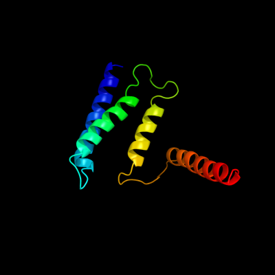
| 1 |
|
PDB 1s7b chain A
Region: 5 - 110
Aligned: 106
Modelled: 106
Confidence: 100.0%
Identity: 100%
Fold: Multidrug resistance efflux transporter EmrE
Superfamily: Multidrug resistance efflux transporter EmrE
Family: Multidrug resistance efflux transporter EmrE
Phyre2
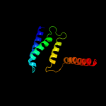
| 2 |
|
PDB 2i68 chain B
Region: 4 - 104
Aligned: 78
Modelled: 101
Confidence: 99.8%
Identity: 99%
PDB header:transport protein
Chain: B: PDB Molecule:protein emre;
PDBTitle: cryo-em based theoretical model structure of transmembrane2 domain of the multidrug-resistance antiporter from e. coli3 emre
Phyre2
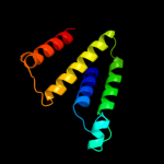
| 3 |
|
PDB 3mp7 chain B
Region: 79 - 101
Aligned: 23
Modelled: 23
Confidence: 17.0%
Identity: 17%
PDB header:protein transport
Chain: B: PDB Molecule:preprotein translocase subunit sece;
PDBTitle: lateral opening of a translocon upon entry of protein suggests the2 mechanism of insertion into membranes
Phyre2
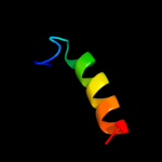
| 4 |
|
PDB 2vqc chain A domain 1
Region: 49 - 70
Aligned: 22
Modelled: 22
Confidence: 10.5%
Identity: 18%
Fold: DNA/RNA-binding 3-helical bundle
Superfamily: "Winged helix" DNA-binding domain
Family: F112-like
Phyre2
| 5 |
|
PDB 2vqc chain A
Region: 49 - 70
Aligned: 22
Modelled: 22
Confidence: 10.5%
Identity: 18%
PDB header:dna-binding protein
Chain: A: PDB Molecule:hypothetical 13.2 kda protein;
PDBTitle: structure of a dna binding winged-helix protein, f-112,2 from sulfolobus spindle-shaped virus 1.
Phyre2
|
| Detailed template information | |
Due to computational demand, binding site predictions are not run for batch jobs
If you want to predict binding sites, please manually submit your model of choice to 3DLigandSite
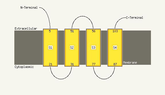
| Transmembrane helix prediction | |
Transmembrane helices have been predicted in your sequence to adopt the topology shown below
Phyre is for academic use only
| Please cite: Protein structure prediction on
the web: a case study using the Phyre server |
| Kelley LA and Sternberg MJE. Nature Protocols
4, 363 - 371 (2009) [pdf] [Import into BibTeX] |
| |
| If you use the binding site
predictions from 3DLigandSite, please also cite: |
| 3DLigandSite: predicting ligand-binding sites using similar structures. |
| Wass MN, Kelley LA and Sternberg
MJ Nucleic Acids Research 38, W469-73 (2010) [PubMed] |
| |
|
|
|
|