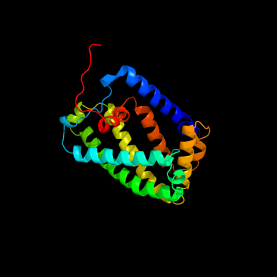
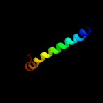
| 1 |
|
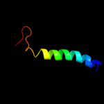
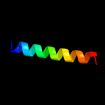
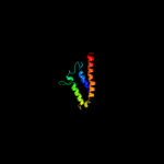
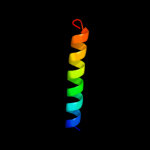
PDB 1y5i chain C domain 1
Region: 2 - 225
Aligned: 216
Modelled: 224
Confidence: 100.0%
Identity: 100%
Fold: Heme-binding four-helical bundle
Superfamily: Respiratory nitrate reductase 1 gamma chain
Family: Respiratory nitrate reductase 1 gamma chain
Phyre2
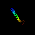
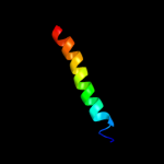
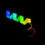
| 2 |
|
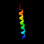
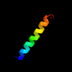
PDB 2knc chain A
Region: 88 - 126
Aligned: 39
Modelled: 39
Confidence: 73.6%
Identity: 26%
PDB header:cell adhesion
Chain: A: PDB Molecule:integrin alpha-iib;
PDBTitle: platelet integrin alfaiib-beta3 transmembrane-cytoplasmic2 heterocomplex
Phyre2
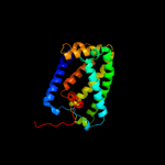
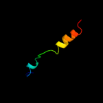
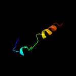
| 3 |
|
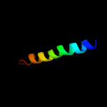
PDB 1kqf chain C
Region: 54 - 199
Aligned: 123
Modelled: 123
Confidence: 67.3%
Identity: 11%
Fold: Heme-binding four-helical bundle
Superfamily: Transmembrane di-heme cytochromes
Family: Formate dehydrogenase N, cytochrome (gamma) subunit
Phyre2
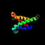
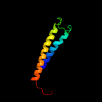
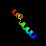
| 4 |
|
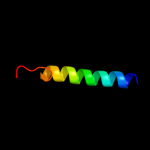
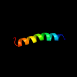
PDB 2klu chain A
Region: 89 - 122
Aligned: 34
Modelled: 34
Confidence: 58.3%
Identity: 26%
PDB header:immune system, membrane protein
Chain: A: PDB Molecule:t-cell surface glycoprotein cd4;
PDBTitle: nmr structure of the transmembrane and cytoplasmic domains2 of human cd4
Phyre2
| 5 |
|
PDB 3kdp chain G
Region: 86 - 112
Aligned: 27
Modelled: 27
Confidence: 55.5%
Identity: 15%
PDB header:hydrolase
Chain: G: PDB Molecule:na+/k+ atpase gamma subunit transcript variant a;
PDBTitle: crystal structure of the sodium-potassium pump
Phyre2
| 6 |
|
PDB 3kdp chain H
Region: 86 - 112
Aligned: 27
Modelled: 27
Confidence: 55.5%
Identity: 15%
PDB header:hydrolase
Chain: H: PDB Molecule:na+/k+ atpase gamma subunit transcript variant a;
PDBTitle: crystal structure of the sodium-potassium pump
Phyre2
| 7 |
|
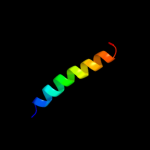
PDB 2k1a chain A
Region: 91 - 119
Aligned: 29
Modelled: 29
Confidence: 46.6%
Identity: 31%
PDB header:cell adhesion
Chain: A: PDB Molecule:integrin alpha-iib;
PDBTitle: bicelle-embedded integrin alpha(iib) transmembrane segment
Phyre2
| 8 |
|
PDB 2jp3 chain A
Region: 86 - 122
Aligned: 37
Modelled: 37
Confidence: 26.5%
Identity: 19%
PDB header:transcription
Chain: A: PDB Molecule:fxyd domain-containing ion transport regulator 4;
PDBTitle: solution structure of the human fxyd4 (chif) protein in sds2 micelles
Phyre2
| 9 |
|
PDB 2jo1 chain A
Region: 86 - 113
Aligned: 28
Modelled: 28
Confidence: 13.3%
Identity: 25%
PDB header:hydrolase regulator
Chain: A: PDB Molecule:phospholemman;
PDBTitle: structure of the na,k-atpase regulatory protein fxyd1 in2 micelles
Phyre2
| 10 |
|
PDB 2k9y chain B
Region: 82 - 112
Aligned: 31
Modelled: 31
Confidence: 9.4%
Identity: 32%
PDB header:transferase
Chain: B: PDB Molecule:ephrin type-a receptor 2;
PDBTitle: epha2 dimeric structure in the lipidic bicelle at ph 5.0
Phyre2
| 11 |
|
PDB 1ar1 chain A
Region: 45 - 122
Aligned: 78
Modelled: 78
Confidence: 9.1%
Identity: 17%
Fold: Cytochrome c oxidase subunit I-like
Superfamily: Cytochrome c oxidase subunit I-like
Family: Cytochrome c oxidase subunit I-like
Phyre2
| 12 |
|
PDB 2zxe chain G
Region: 86 - 109
Aligned: 24
Modelled: 24
Confidence: 7.1%
Identity: 29%
PDB header:hydrolase/transport protein
Chain: G: PDB Molecule:phospholemman-like protein;
PDBTitle: crystal structure of the sodium - potassium pump in the e2.2k+.pi2 state
Phyre2
| 13 |
|
PDB 2k9y chain A
Region: 82 - 112
Aligned: 31
Modelled: 31
Confidence: 7.0%
Identity: 32%
PDB header:transferase
Chain: A: PDB Molecule:ephrin type-a receptor 2;
PDBTitle: epha2 dimeric structure in the lipidic bicelle at ph 5.0
Phyre2
| 14 |
|
PDB 3rko chain A
Region: 17 - 36
Aligned: 20
Modelled: 20
Confidence: 5.7%
Identity: 25%
PDB header:oxidoreductase
Chain: A: PDB Molecule:nadh-quinone oxidoreductase subunit a;
PDBTitle: crystal structure of the membrane domain of respiratory complex i from2 e. coli at 3.0 angstrom resolution
Phyre2
| 15 |
|
PDB 2ww9 chain B
Region: 187 - 214
Aligned: 25
Modelled: 28
Confidence: 5.7%
Identity: 28%
PDB header:ribosome
Chain: B: PDB Molecule:protein transport protein sss1;
PDBTitle: cryo-em structure of the active yeast ssh1 complex bound to the2 yeast 80s ribosome
Phyre2
| 16 |
|
PDB 1wpg chain A domain 4
Region: 15 - 115
Aligned: 94
Modelled: 101
Confidence: 5.4%
Identity: 12%
Fold: Calcium ATPase, transmembrane domain M
Superfamily: Calcium ATPase, transmembrane domain M
Family: Calcium ATPase, transmembrane domain M
Phyre2
| 17 |
|
PDB 2k1k chain B
Region: 86 - 112
Aligned: 27
Modelled: 27
Confidence: 5.3%
Identity: 26%
PDB header:signaling protein
Chain: B: PDB Molecule:ephrin type-a receptor 1;
PDBTitle: nmr structures of dimeric transmembrane domain of the2 receptor tyrosine kinase epha1 in lipid bicelles at ph 4.3
Phyre2
| 18 |
|
PDB 2k1l chain A
Region: 86 - 112
Aligned: 27
Modelled: 27
Confidence: 5.3%
Identity: 26%
PDB header:signaling protein
Chain: A: PDB Molecule:ephrin type-a receptor 1;
PDBTitle: nmr structures of dimeric transmembrane domain of the2 receptor tyrosine kinase epha1 in lipid bicelles at ph 6.3
Phyre2
| 19 |
|
PDB 2k1l chain B
Region: 86 - 112
Aligned: 27
Modelled: 27
Confidence: 5.3%
Identity: 26%
PDB header:signaling protein
Chain: B: PDB Molecule:ephrin type-a receptor 1;
PDBTitle: nmr structures of dimeric transmembrane domain of the2 receptor tyrosine kinase epha1 in lipid bicelles at ph 6.3
Phyre2
| 20 |
|
PDB 2k1k chain A
Region: 86 - 112
Aligned: 27
Modelled: 27
Confidence: 5.3%
Identity: 26%
PDB header:signaling protein
Chain: A: PDB Molecule:ephrin type-a receptor 1;
PDBTitle: nmr structures of dimeric transmembrane domain of the2 receptor tyrosine kinase epha1 in lipid bicelles at ph 4.3
Phyre2
| 21 |
|