| 1 |
|
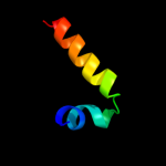
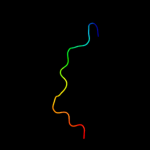
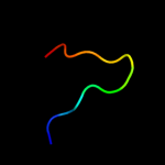
PDB 2hzq chain A
Region: 8 - 25
Aligned: 18
Modelled: 18
Confidence: 17.6%
Identity: 33%
PDB header:transport protein
Chain: A: PDB Molecule:apolipoprotein d;
PDBTitle: crystal structure of human apolipoprotein d (apod) in2 complex with progesterone
Phyre2
| 2 |
|
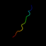
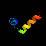
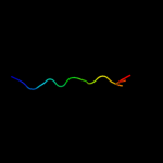
PDB 1vd3 chain A
Region: 3 - 32
Aligned: 30
Modelled: 30
Confidence: 14.2%
Identity: 40%
PDB header:hydrolase
Chain: A: PDB Molecule:rnase ngr3;
PDBTitle: ribonuclease nt in complex with 2'-ump
Phyre2
| 3 |
|
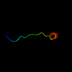
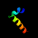
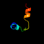
PDB 1iyb chain A
Region: 3 - 32
Aligned: 30
Modelled: 30
Confidence: 13.8%
Identity: 37%
Fold: Ribonuclease Rh-like
Superfamily: Ribonuclease Rh-like
Family: Ribonuclease Rh-like
Phyre2
| 4 |
|
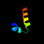
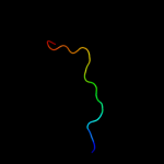
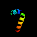
PDB 2pqx chain A
Region: 3 - 32
Aligned: 30
Modelled: 30
Confidence: 11.2%
Identity: 17%
PDB header:hydrolase
Chain: A: PDB Molecule:ribonuclease i;
PDBTitle: e. coli rnase 1 (in vivo folded)
Phyre2
| 5 |
|
PDB 1kxo chain A
Region: 10 - 24
Aligned: 15
Modelled: 15
Confidence: 10.5%
Identity: 27%
Fold: Lipocalins
Superfamily: Lipocalins
Family: Retinol binding protein-like
Phyre2
| 6 |
|
PDB 1bbp chain A
Region: 8 - 27
Aligned: 19
Modelled: 20
Confidence: 10.2%
Identity: 26%
Fold: Lipocalins
Superfamily: Lipocalins
Family: Retinol binding protein-like
Phyre2
| 7 |
|
PDB 1dix chain A
Region: 3 - 32
Aligned: 30
Modelled: 30
Confidence: 9.1%
Identity: 30%
Fold: Ribonuclease Rh-like
Superfamily: Ribonuclease Rh-like
Family: Ribonuclease Rh-like
Phyre2
| 8 |
|
PDB 1n0s chain A
Region: 8 - 24
Aligned: 17
Modelled: 17
Confidence: 8.4%
Identity: 29%
Fold: Lipocalins
Superfamily: Lipocalins
Family: Retinol binding protein-like
Phyre2
| 9 |
|
PDB 1sgl chain A
Region: 3 - 32
Aligned: 30
Modelled: 30
Confidence: 8.1%
Identity: 23%
Fold: Ribonuclease Rh-like
Superfamily: Ribonuclease Rh-like
Family: Ribonuclease Rh-like
Phyre2
| 10 |
|
PDB 1jy5 chain A
Region: 3 - 32
Aligned: 30
Modelled: 30
Confidence: 7.5%
Identity: 23%
Fold: Ribonuclease Rh-like
Superfamily: Ribonuclease Rh-like
Family: Ribonuclease Rh-like
Phyre2
| 11 |
|
PDB 1z24 chain A domain 1
Region: 9 - 25
Aligned: 17
Modelled: 17
Confidence: 7.4%
Identity: 18%
Fold: Lipocalins
Superfamily: Lipocalins
Family: Retinol binding protein-like
Phyre2
| 12 |
|
PDB 1sg1 chain X domain 2
Region: 115 - 127
Aligned: 13
Modelled: 13
Confidence: 6.9%
Identity: 46%
Fold: TNF receptor-like
Superfamily: TNF receptor-like
Family: TNF receptor-like
Phyre2
| 13 |
|
PDB 1gka chain B
Region: 9 - 24
Aligned: 16
Modelled: 16
Confidence: 5.9%
Identity: 31%
Fold: Lipocalins
Superfamily: Lipocalins
Family: Retinol binding protein-like
Phyre2
| 14 |
|
PDB 1ucd chain A
Region: 3 - 32
Aligned: 30
Modelled: 30
Confidence: 5.9%
Identity: 30%
Fold: Ribonuclease Rh-like
Superfamily: Ribonuclease Rh-like
Family: Ribonuclease Rh-like
Phyre2
| 15 |
|
PDB 3d3z chain A
Region: 3 - 32
Aligned: 30
Modelled: 30
Confidence: 5.6%
Identity: 27%
PDB header:hydrolase
Chain: A: PDB Molecule:actibind;
PDBTitle: crystal structure of actibind a t2 rnase
Phyre2
| 16 |
|
PDB 2a0s chain A domain 1
Region: 4 - 15
Aligned: 12
Modelled: 12
Confidence: 5.5%
Identity: 25%
Fold: T-fold
Superfamily: Tetrahydrobiopterin biosynthesis enzymes-like
Family: 6-pyruvoyl tetrahydropterin synthase
Phyre2
| 17 |
|
PDB 1y13 chain A
Region: 4 - 15
Aligned: 12
Modelled: 12
Confidence: 5.5%
Identity: 25%
Fold: T-fold
Superfamily: Tetrahydrobiopterin biosynthesis enzymes-like
Family: 6-pyruvoyl tetrahydropterin synthase
Phyre2