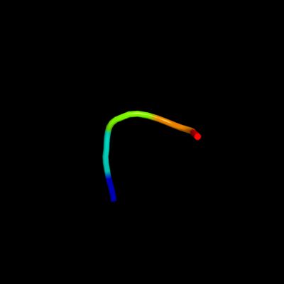
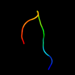
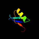
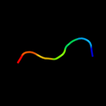
Phyre Home Retrieve Phyre Job Id
Emaill.a.kelley@imperial.ac.uk
DescriptionP29010
DateWed Jan 25 15:20:47 GMT 2012
Unique Job ID8ddc8f202c6cde4d

Summary 

Top model
Image coloured by rainbow N → C terminus
Model (left) based on template c1wqkA_
Top template information
PDB header:toxin
Chain: A: PDB Molecule:toxin apetx1;
PDBTitle: solution structure of apetx1, a specific peptide inhibitor2 of human ether-a-go-go-related gene potassium channels3 from the venom of the sea anemone anthopleura4 elegantissima: a new fold for an herg toxin
Confidence and coverage
Confidence: 15.9% Coverage: 8%
5 residues ( 8% of your sequence) have been modelled with 15.9% confidence by the single highest scoring template.
You may wish to submit your sequence to Phyrealarm. This will automatically scan your sequence every week for new potential templates as they appear in the Phyre2 library.
Please note: You must be registered and logged in to use Phyrealarm.
3D viewing
Interactive 3D view in Jmol

Sequence analysis 

Secondary structure and disorder prediction 

   1........10.........20.........30.........40.........50.........60
Sequence  MNSAFVLVLTVFLVSGEPVDIAVSVHRTMQECMTAATEQKIPGNCYPVDKVIHQDNIEIP
Secondary structure 


SS confidence 


Disorder  ?????


??????
??


????
Disorder confidence 


   ...
Sequence  AGL
Secondary structure 


SS confidence 


Disorder  ???
Disorder confidence 


Confidence Key
High(9)                    Low (0)
?Disordered
Alpha helix
Beta strand

Domain analysis 

Hover over an aligned region to see model and summary info

Please note, only up to the top 20 hits are modelled to reduce computer load

RankAligned region

PDB 1wqk chain A

3D model

Region: 45 - 49
Aligned: 5
Modelled: 5
Confidence: 15.9%
Identity: 100%
PDB header:toxin
Chain: A: PDB Molecule:toxin apetx1;
PDBTitle: solution structure of apetx1, a specific peptide inhibitor2 of human ether-a-go-go-related gene potassium channels3 from the venom of the sea anemone anthopleura4 elegantissima: a new fold for an herg toxin

Phyre2

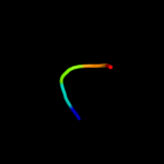
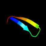
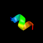
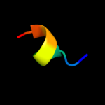
PDB 2guy chain A domain 1

3D model

Region: 9 - 19
Aligned: 11
Modelled: 11
Confidence: 11.3%
Identity: 27%
Fold: Glycosyl hydrolase domain
Superfamily: Glycosyl hydrolase domain
Family: alpha-Amylases, C-terminal beta-sheet domain

Phyre2

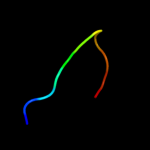
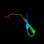
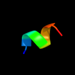
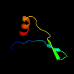
PDB 2aaa chain A domain 1

3D model

Region: 9 - 19
Aligned: 11
Modelled: 11
Confidence: 10.2%
Identity: 36%
Fold: Glycosyl hydrolase domain
Superfamily: Glycosyl hydrolase domain
Family: alpha-Amylases, C-terminal beta-sheet domain

Phyre2

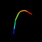
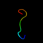
PDB 2taa chain A domain 1

3D model

Region: 9 - 19
Aligned: 11
Modelled: 11
Confidence: 8.7%
Identity: 27%
Fold: Glycosyl hydrolase domain
Superfamily: Glycosyl hydrolase domain
Family: alpha-Amylases, C-terminal beta-sheet domain

Phyre2

PDB 2qtx chain L

3D model

Region: 9 - 23
Aligned: 15
Modelled: 15
Confidence: 8.5%
Identity: 40%
PDB header:rna binding protein
Chain: L: PDB Molecule:uncharacterized protein mj1435;
PDBTitle: crystal structure of an hfq-like protein from methanococcus2 jannaschii

Phyre2

PDB 1kq1 chain W

3D model

Region: 9 - 28
Aligned: 20
Modelled: 20
Confidence: 7.0%
Identity: 20%
PDB header:translation
Chain: W: PDB Molecule:host factor for q beta;
PDBTitle: 1.55 a crystal structure of the pleiotropic translational2 regulator, hfq

Phyre2

PDB 1wxn chain A

3D model

Region: 45 - 49
Aligned: 5
Modelled: 5
Confidence: 6.4%
Identity: 60%
PDB header:toxin
Chain: A: PDB Molecule:toxin apetx2;
PDBTitle: solution structure of apetx2, a specific peptide inhibitor2 of asic3 proton-gated channels

Phyre2

PDB 2qsd chain B

3D model

Region: 11 - 51
Aligned: 41
Modelled: 41
Confidence: 6.3%
Identity: 27%
PDB header:structural genomics, unknown function
Chain: B: PDB Molecule:uncharacterized conserved protein;
PDBTitle: crystal structure of a protein il1583 from idiomarina loihiensis

Phyre2

PDB 2hwn chain F

3D model

Region: 25 - 32
Aligned: 8
Modelled: 8
Confidence: 6.0%
Identity: 50%
PDB header:transferase
Chain: F: PDB Molecule:a kinase binding peptide;
PDBTitle: crystal structure of rii alpha dimerization/docking domain of pka2 bound to the d-akap2 peptide

Phyre2

PDB 2hwn chain E

3D model

Region: 25 - 32
Aligned: 8
Modelled: 8
Confidence: 6.0%
Identity: 50%
PDB header:transferase
Chain: E: PDB Molecule:a kinase binding peptide;
PDBTitle: crystal structure of rii alpha dimerization/docking domain of pka2 bound to the d-akap2 peptide

Phyre2

PDB 2qam chain C domain 1

3D model

Region: 42 - 51
Aligned: 10
Modelled: 10
Confidence: 6.0%
Identity: 40%
Fold: SH3-like barrel
Superfamily: Translation proteins SH3-like domain
Family: C-terminal domain of ribosomal protein L2

Phyre2

PDB 1p9q chain C domain 3

3D model

Region: 57 - 63
Aligned: 7
Modelled: 7
Confidence: 5.9%
Identity: 43%
Fold: Ferredoxin-like
Superfamily: EF-G C-terminal domain-like
Family: Hypothetical protein AF0491, C-terminal domain

Phyre2

PDB 1qf8 chain A

3D model

Region: 50 - 63
Aligned: 14
Modelled: 14
Confidence: 5.9%
Identity: 29%
Fold: Rubredoxin-like
Superfamily: Casein kinase II beta subunit
Family: Casein kinase II beta subunit

Phyre2

PDB 1u1s chain A domain 1

3D model

Region: 9 - 28
Aligned: 20
Modelled: 20
Confidence: 5.6%
Identity: 15%
Fold: Sm-like fold
Superfamily: Sm-like ribonucleoproteins
Family: Pleiotropic translational regulator Hfq

Phyre2

PDB 1t95 chain A domain 3

3D model

Region: 57 - 63
Aligned: 7
Modelled: 5
Confidence: 5.6%
Identity: 43%
Fold: Ferredoxin-like
Superfamily: EF-G C-terminal domain-like
Family: Hypothetical protein AF0491, C-terminal domain

Phyre2

PDB 1w7v chain D

3D model

Region: 12 - 44
Aligned: 33
Modelled: 33
Confidence: 5.4%
Identity: 18%
PDB header:hydrolase
Chain: D: PDB Molecule:xaa-pro aminopeptidase;
PDBTitle: znmg substituted aminopeptidase p from e. coli

Phyre2

PDB 2aiv chain A

3D model

Region: 44 - 50
Aligned: 7
Modelled: 7
Confidence: 5.4%
Identity: 100%
PDB header:transport protein
Chain: A: PDB Molecule:fragment of nucleoporin nup116/nsp116;
PDBTitle: multiple conformations in the ligand-binding site of the2 yeast nuclear pore targeting domain of nup116p

Phyre2

PDB 2guk chain A domain 1

3D model

Region: 26 - 33
Aligned: 8
Modelled: 8
Confidence: 5.2%
Identity: 13%
Fold: PG1857-like
Superfamily: PG1857-like
Family: PG1857-like

Phyre2

PDB 2i9x chain A domain 1

3D model

Region: 2 - 36
Aligned: 35
Modelled: 35
Confidence: 5.0%
Identity: 23%
Fold: SpoVG-like
Superfamily: SpoVG-like
Family: SpoVG-like

Phyre2
1

c1wqkA_
2

d2guya1
3

d2aaaa1
4

d2taaa1
5

c2qtxL_
6

c1kq1W_
7

c1wxnA_
8

c2qsdB_
9

c2hwnF_
10

c2hwnE_
11

d2qamc1
12

d1p9qc3
13

d1qf8a_
14

d1u1sa1
15

d1t95a3
16

c1w7vD_
17

c2aivA_
18

d2guka1
19

d2i9xa1


Detailed template information 

#
Template Alignment Coverage3D Model Confidence
% i.d. Template Information
1c1wqkA_


15.9 100 PDB header:toxin
Chain: A: PDB Molecule:toxin apetx1;
PDBTitle: solution structure of apetx1, a specific peptide inhibitor2 of human ether-a-go-go-related gene potassium channels3 from the venom of the sea anemone anthopleura4 elegantissima: a new fold for an herg toxin
2d2guya1


11.3 27 Fold:Glycosyl hydrolase domain
Superfamily:Glycosyl hydrolase domain
Family:alpha-Amylases, C-terminal beta-sheet domain
3d2aaaa1


10.2 36 Fold:Glycosyl hydrolase domain
Superfamily:Glycosyl hydrolase domain
Family:alpha-Amylases, C-terminal beta-sheet domain
4d2taaa1


8.7 27 Fold:Glycosyl hydrolase domain
Superfamily:Glycosyl hydrolase domain
Family:alpha-Amylases, C-terminal beta-sheet domain
5c2qtxL_


8.5 40 PDB header:rna binding protein
Chain: L: PDB Molecule:uncharacterized protein mj1435;
PDBTitle: crystal structure of an hfq-like protein from methanococcus2 jannaschii
6c1kq1W_


7.0 20 PDB header:translation
Chain: W: PDB Molecule:host factor for q beta;
PDBTitle: 1.55 a crystal structure of the pleiotropic translational2 regulator, hfq
7c1wxnA_


6.4 60 PDB header:toxin
Chain: A: PDB Molecule:toxin apetx2;
PDBTitle: solution structure of apetx2, a specific peptide inhibitor2 of asic3 proton-gated channels
8c2qsdB_


6.3 27 PDB header:structural genomics, unknown function
Chain: B: PDB Molecule:uncharacterized conserved protein;
PDBTitle: crystal structure of a protein il1583 from idiomarina loihiensis
9c2hwnF_


6.0 50 PDB header:transferase
Chain: F: PDB Molecule:a kinase binding peptide;
PDBTitle: crystal structure of rii alpha dimerization/docking domain of pka2 bound to the d-akap2 peptide
10c2hwnE_


6.0 50 PDB header:transferase
Chain: E: PDB Molecule:a kinase binding peptide;
PDBTitle: crystal structure of rii alpha dimerization/docking domain of pka2 bound to the d-akap2 peptide
11d2qamc1


6.0 40 Fold:SH3-like barrel
Superfamily:Translation proteins SH3-like domain
Family:C-terminal domain of ribosomal protein L2
12d1p9qc3


5.9 43 Fold:Ferredoxin-like
Superfamily:EF-G C-terminal domain-like
Family:Hypothetical protein AF0491, C-terminal domain
13d1qf8a_


5.9 29 Fold:Rubredoxin-like
Superfamily:Casein kinase II beta subunit
Family:Casein kinase II beta subunit
14d1u1sa1


5.6 15 Fold:Sm-like fold
Superfamily:Sm-like ribonucleoproteins
Family:Pleiotropic translational regulator Hfq
15d1t95a3


5.6 43 Fold:Ferredoxin-like
Superfamily:EF-G C-terminal domain-like
Family:Hypothetical protein AF0491, C-terminal domain
16c1w7vD_


5.4 18 PDB header:hydrolase
Chain: D: PDB Molecule:xaa-pro aminopeptidase;
PDBTitle: znmg substituted aminopeptidase p from e. coli
17c2aivA_


5.4 100 PDB header:transport protein
Chain: A: PDB Molecule:fragment of nucleoporin nup116/nsp116;
PDBTitle: multiple conformations in the ligand-binding site of the2 yeast nuclear pore targeting domain of nup116p
18d2guka1


5.2 13 Fold:PG1857-like
Superfamily:PG1857-like
Family:PG1857-like
19d2i9xa1


5.0 23 Fold:SpoVG-like
Superfamily:SpoVG-like
Family:SpoVG-like

Binding site prediction 

Due to computational demand, binding site predictions are not run for batch jobs

If you want to predict binding sites, please manually submit your model of choice to 3DLigandSite


Phyre is for academic use only

Please cite: Protein structure prediction on the web: a case study using the Phyre server
Kelley LA and Sternberg MJE. Nature Protocols 4, 363 - 371 (2009) [pdf] [Import into BibTeX]
 
If you use the binding site predictions from 3DLigandSite, please also cite:
3DLigandSite: predicting ligand-binding sites using similar structures.
Wass MN, Kelley LA and Sternberg MJ Nucleic Acids Research 38, W469-73 (2010) [PubMed]
 
© Structural Bioinformatics Group
Imperial College London
Lawrence Kelley, Benjamin Jefferys
Disclaimer
Terms and Conditions
Component software
Template detection: HHpred 1.51
Secondary structure prediction: Psi-pred 2.5
Disorder prediction: Disopred 2.4
Transmembrane prediction: Memsat_SVM
Multi-template modelling and ab initio: Poing 1.0