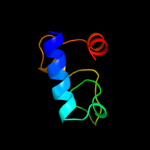
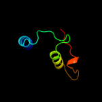
| 1 |
|
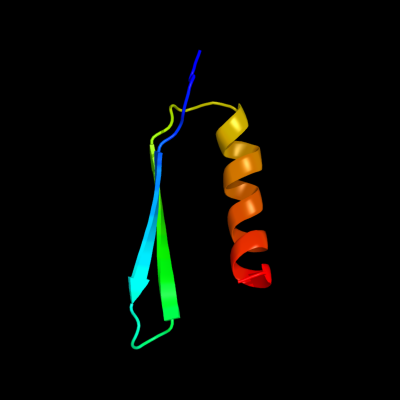
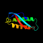
PDB 2rrl chain A
Region: 58 - 101
Aligned: 42
Modelled: 44
Confidence: 60.7%
Identity: 21%
PDB header:protein transport
Chain: A: PDB Molecule:flagellar hook-length control protein;
PDBTitle: solution structure of the c-terminal domain of the flik
Phyre2
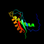
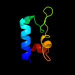
| 2 |
|
PDB 2rpb chain A
Region: 57 - 151
Aligned: 91
Modelled: 95
Confidence: 42.9%
Identity: 11%
PDB header:membrane protein
Chain: A: PDB Molecule:hypothetical membrane protein;
PDBTitle: the solution structure of membrane protein
Phyre2
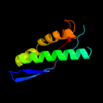
| 3 |
|
PDB 3bk6 chain C
Region: 57 - 151
Aligned: 91
Modelled: 95
Confidence: 42.7%
Identity: 10%
PDB header:membrane protein
Chain: C: PDB Molecule:ph stomatin;
PDBTitle: crystal structure of a core domain of stomatin from2 pyrococcus horikoshii
Phyre2
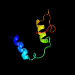
| 4 |
|
PDB 3gxq chain B
Region: 95 - 126
Aligned: 31
Modelled: 32
Confidence: 33.3%
Identity: 13%
PDB header:dna binding protein/dna
Chain: B: PDB Molecule:putative regulator of transfer genes arta;
PDBTitle: structure of arta and dna complex
Phyre2
| 5 |
|
PDB 3u5g chain B
Region: 62 - 150
Aligned: 81
Modelled: 88
Confidence: 19.7%
Identity: 19%
PDB header:ribosome
Chain: B: PDB Molecule:40s ribosomal protein s1-a;
PDBTitle: the structure of the eukaryotic ribosome at 3.0 a resolution
Phyre2
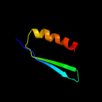
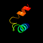
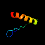
| 6 |
|
PDB 1win chain A
Region: 41 - 151
Aligned: 107
Modelled: 111
Confidence: 13.2%
Identity: 7%
Fold: EF-Ts domain-like
Superfamily: Band 7/SPFH domain
Family: Band 7/SPFH domain
Phyre2
| 7 |
|
PDB 2xzm chain 4
Region: 62 - 150
Aligned: 81
Modelled: 89
Confidence: 11.0%
Identity: 9%
PDB header:ribosome
Chain: 4: PDB Molecule:40s ribosomal protein s3a;
PDBTitle: crystal structure of the eukaryotic 40s ribosomal2 subunit in complex with initiation factor 1. this file3 contains the 40s subunit and initiation factor for4 molecule 1
Phyre2
| 8 |
|
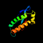
PDB 2od5 chain A domain 1
Region: 93 - 147
Aligned: 44
Modelled: 55
Confidence: 7.5%
Identity: 20%
Fold: DNA/RNA-binding 3-helical bundle
Superfamily: "Winged helix" DNA-binding domain
Family: Marine metagenome family WH1
Phyre2
| 9 |
|
PDB 2od5 chain A
Region: 93 - 147
Aligned: 44
Modelled: 55
Confidence: 7.5%
Identity: 20%
PDB header:dna binding protein
Chain: A: PDB Molecule:hypothetical protein;
PDBTitle: crystal structure of a putative nucleic acid binding protein2 (jcvi_pep_1096688149193) from uncultured marine organism at 1.79 a3 resolution
Phyre2
| 10 |
|
PDB 3dyd chain B
Region: 95 - 150
Aligned: 52
Modelled: 52
Confidence: 7.1%
Identity: 21%
PDB header:transferase
Chain: B: PDB Molecule:tyrosine aminotransferase;
PDBTitle: human tyrosine aminotransferase
Phyre2
| 11 |
|
PDB 2dyj chain A domain 1
Region: 64 - 101
Aligned: 38
Modelled: 38
Confidence: 6.1%
Identity: 21%
Fold: Alpha-lytic protease prodomain-like
Superfamily: Ribosome-binding factor A, RbfA
Family: Ribosome-binding factor A, RbfA
Phyre2
| 12 |
|
PDB 3pdx chain A
Region: 95 - 150
Aligned: 52
Modelled: 52
Confidence: 5.5%
Identity: 19%
PDB header:transferase
Chain: A: PDB Molecule:tyrosine aminotransferase;
PDBTitle: crystal structural of mouse tyrosine aminotransferase
Phyre2