Phyre Home Retrieve Phyre Job Id
Emaill.a.kelley@imperial.ac.uk
DescriptionP76069
DateThu Jan 5 12:18:08 GMT 2012
Unique Job ID8820b6e5671fd242

Summary 

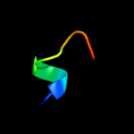
Top model
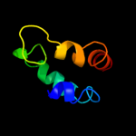
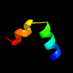
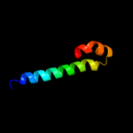
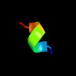
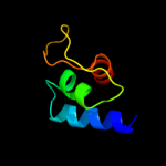
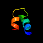
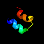
Image coloured by rainbow N → C terminus
Model (left) based on template d2hrkb1
Top template information
Fold:GST C-terminal domain-like
Superfamily:GST C-terminal domain-like
Family:Arc1p N-terminal domain-like
Confidence and coverage
Confidence: 39.4% Coverage: 43%
51 residues ( 43% of your sequence) have been modelled with 39.4% confidence by the single highest scoring template.
You may wish to submit your sequence to Phyrealarm. This will automatically scan your sequence every week for new potential templates as they appear in the Phyre2 library.
Please note: You must be registered and logged in to use Phyrealarm.
3D viewing
Interactive 3D view in Jmol

Sequence analysis 

Secondary structure and disorder prediction 

   1........10.........20.........30.........40.........50.........60
Sequence  MSRSSDNDQYRSRNALIRRHIEKMDASLHVGTKEFDISKVSEVDSVDDLLIDNAARYLLK
Secondary structure 


SS confidence 


Disorder  ???????


???????
??????


Disorder confidence 


   .........70.........80.........90.........100.........110.........
Sequence  DWKGVGELVNGVEVALEYTAERGIALLKQNPELYWQILAEAASIAQGKEQQKQDTIKKP
Secondary structure 


SS confidence 


Disorder 


??????????


????
Disorder confidence 


Confidence Key
High(9)                    Low (0)
?Disordered
Alpha helix
Beta strand

Domain analysis 

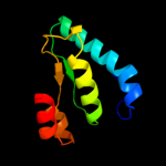
Hover over an aligned region to see model and summary info

Please note, only up to the top 20 hits are modelled to reduce computer load

RankAligned region

PDB 2hrk chain B domain 1

3D model

Region: 8 - 62
Aligned: 51
Modelled: 55
Confidence: 39.4%
Identity: 22%
Fold: GST C-terminal domain-like
Superfamily: GST C-terminal domain-like
Family: Arc1p N-terminal domain-like

Phyre2

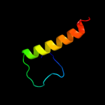
PDB 3hdf chain A

3D model

Region: 64 - 101
Aligned: 38
Modelled: 38
Confidence: 24.4%
Identity: 16%
PDB header:hydrolase
Chain: A: PDB Molecule:lysozyme;
PDBTitle: crystal structure of truncated endolysin r21 from phage 21

Phyre2

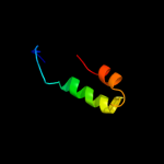
PDB 2elk chain A

3D model

Region: 31 - 71
Aligned: 41
Modelled: 41
Confidence: 17.8%
Identity: 22%
PDB header:transcription
Chain: A: PDB Molecule:spcc24b10.08c protein;
PDBTitle: solution structure of the sant domain of fission yeast2 spcc24b10.08c protein

Phyre2

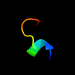
PDB 2dii chain A

3D model

Region: 85 - 94
Aligned: 10
Modelled: 10
Confidence: 15.4%
Identity: 30%
PDB header:transcription
Chain: A: PDB Molecule:tfiih basal transcription factor complex p62
PDBTitle: solution structure of the bsd domain of human tfiih basal2 transcription factor complex p62 subunit

Phyre2

PDB 2dii chain A domain 1

3D model

Region: 85 - 94
Aligned: 10
Modelled: 10
Confidence: 15.0%
Identity: 30%
Fold: BSD domain-like
Superfamily: BSD domain-like
Family: BSD domain

Phyre2

PDB 3e2s chain A

3D model

Region: 1 - 88
Aligned: 85
Modelled: 88
Confidence: 15.0%
Identity: 13%
PDB header:oxidoreductase
Chain: A: PDB Molecule:proline dehydrogenase;
PDBTitle: crystal structure reduced puta86-630 mutant y540s complexed with l-2 proline

Phyre2

PDB 1nh1 chain A

3D model

Region: 37 - 109
Aligned: 68
Modelled: 68
Confidence: 14.7%
Identity: 16%
Fold: Antivirulence factor
Superfamily: Antivirulence factor
Family: Antivirulence factor

Phyre2

PDB 1nh1 chain A

3D model

Region: 37 - 109
Aligned: 68
Modelled: 68
Confidence: 14.7%
Identity: 16%
PDB header:avirulence protein
Chain: A: PDB Molecule:avirulence b protein;
PDBTitle: crystal structure of the type iii effector avrb from2 pseudomonas syringae.

Phyre2

PDB 2l9b chain A

3D model

Region: 79 - 99
Aligned: 21
Modelled: 21
Confidence: 14.1%
Identity: 24%
PDB header:transcription
Chain: A: PDB Molecule:mrna 3'-end-processing protein rna15;
PDBTitle: heterodimer between rna14p monkeytail domain and rna15p hinge domain2 of the yeast cf ia complex

Phyre2

PDB 1gvd chain A

3D model

Region: 47 - 71
Aligned: 25
Modelled: 25
Confidence: 13.9%
Identity: 28%
Fold: DNA/RNA-binding 3-helical bundle
Superfamily: Homeodomain-like
Family: Myb/SANT domain

Phyre2

PDB 1x41 chain A domain 1

3D model

Region: 47 - 71
Aligned: 25
Modelled: 25
Confidence: 13.7%
Identity: 12%
Fold: DNA/RNA-binding 3-helical bundle
Superfamily: Homeodomain-like
Family: Homeodomain

Phyre2

PDB 1h88 chain C

3D model

Region: 47 - 71
Aligned: 25
Modelled: 25
Confidence: 13.0%
Identity: 20%
PDB header:transcription/dna
Chain: C: PDB Molecule:myb proto-oncogene protein;
PDBTitle: crystal structure of ternary protein-dna complex1

Phyre2

PDB 2oku chain A domain 1

3D model

Region: 8 - 59
Aligned: 41
Modelled: 41
Confidence: 11.5%
Identity: 22%
Fold: N-cbl like
Superfamily: PG0775 C-terminal domain-like
Family: PG0775 C-terminal domain-like

Phyre2

PDB 2dim chain A

3D model

Region: 31 - 71
Aligned: 41
Modelled: 41
Confidence: 11.4%
Identity: 15%
PDB header:dna binding protein
Chain: A: PDB Molecule:cell division cycle 5-like protein;
PDBTitle: solution structure of the myb_dna-binding domain of human2 cell division cycle 5-like protein

Phyre2

PDB 1wxp chain A

3D model

Region: 31 - 66
Aligned: 36
Modelled: 36
Confidence: 11.0%
Identity: 22%
PDB header:transport protein
Chain: A: PDB Molecule:tho complex subunit 1;
PDBTitle: solution structure of the death domain of nuclear matrix2 protein p84

Phyre2

PDB 1a5j chain A domain 1

3D model

Region: 47 - 71
Aligned: 25
Modelled: 25
Confidence: 10.6%
Identity: 24%
Fold: DNA/RNA-binding 3-helical bundle
Superfamily: Homeodomain-like
Family: Myb/SANT domain

Phyre2

PDB 2elp chain A

3D model

Region: 17 - 23
Aligned: 7
Modelled: 7
Confidence: 10.1%
Identity: 43%
PDB header:transcription
Chain: A: PDB Molecule:zinc finger protein 406;
PDBTitle: solution structure of the 13th c2h2 zinc finger of human2 zinc finger protein 406

Phyre2

PDB 4a69 chain C

3D model

Region: 43 - 96
Aligned: 40
Modelled: 54
Confidence: 10.0%
Identity: 8%
PDB header:transcription
Chain: C: PDB Molecule:nuclear receptor corepressor 2;
PDBTitle: structure of hdac3 bound to corepressor and inositol tetraphosphate

Phyre2

PDB 2yus chain A

3D model

Region: 47 - 71
Aligned: 24
Modelled: 25
Confidence: 9.9%
Identity: 33%
PDB header:transcription
Chain: A: PDB Molecule:swi/snf-related matrix-associated actin-
PDBTitle: solution structure of the sant domain of human swi/snf-2 related matrix-associated actin-dependent regulator of3 chromatin subfamily c member 1

Phyre2

PDB 1x41 chain A

3D model

Region: 47 - 71
Aligned: 25
Modelled: 25
Confidence: 9.8%
Identity: 12%
PDB header:transcription
Chain: A: PDB Molecule:transcriptional adaptor 2-like, isoform b;
PDBTitle: solution structure of the myb-like dna binding domain of2 human transcriptional adaptor 2-like, isoform b

Phyre2
1

d2hrkb1
2

c3hdfA_
3

c2elkA_
4

c2diiA_
5

d2diia1
6

c3e2sA_
7

d1nh1a_
8

c1nh1A_
9

c2l9bA_
10

d1gvda_
11

d1x41a1
12

c1h88C_
13

d2okua1
14

c2dimA_
15

c1wxpA_
16

d1a5ja1
17

c2elpA_
18

c4a69C_
19

c2yusA_
20

c1x41A_
21


22


23


24


25


26


27


28


29


30


31


32


33


34


35


36


37


38


39


40


41


42


43


44


45


46


47


48


49


Detailed template information 

#
Template Alignment Coverage3D Model Confidence
% i.d. Template Information
1d2hrkb1


39.4 22 Fold:GST C-terminal domain-like
Superfamily:GST C-terminal domain-like
Family:Arc1p N-terminal domain-like
2c3hdfA_


24.4 16 PDB header:hydrolase
Chain: A: PDB Molecule:lysozyme;
PDBTitle: crystal structure of truncated endolysin r21 from phage 21
3c2elkA_


17.8 22 PDB header:transcription
Chain: A: PDB Molecule:spcc24b10.08c protein;
PDBTitle: solution structure of the sant domain of fission yeast2 spcc24b10.08c protein
4c2diiA_


15.4 30 PDB header:transcription
Chain: A: PDB Molecule:tfiih basal transcription factor complex p62
PDBTitle: solution structure of the bsd domain of human tfiih basal2 transcription factor complex p62 subunit
5d2diia1


15.0 30 Fold:BSD domain-like
Superfamily:BSD domain-like
Family:BSD domain
6c3e2sA_


15.0 13 PDB header:oxidoreductase
Chain: A: PDB Molecule:proline dehydrogenase;
PDBTitle: crystal structure reduced puta86-630 mutant y540s complexed with l-2 proline
7d1nh1a_


14.7 16 Fold:Antivirulence factor
Superfamily:Antivirulence factor
Family:Antivirulence factor
8c1nh1A_


14.7 16 PDB header:avirulence protein
Chain: A: PDB Molecule:avirulence b protein;
PDBTitle: crystal structure of the type iii effector avrb from2 pseudomonas syringae.
9c2l9bA_


14.1 24 PDB header:transcription
Chain: A: PDB Molecule:mrna 3'-end-processing protein rna15;
PDBTitle: heterodimer between rna14p monkeytail domain and rna15p hinge domain2 of the yeast cf ia complex
10d1gvda_


13.9 28 Fold:DNA/RNA-binding 3-helical bundle
Superfamily:Homeodomain-like
Family:Myb/SANT domain
11d1x41a1


13.7 12 Fold:DNA/RNA-binding 3-helical bundle
Superfamily:Homeodomain-like
Family:Homeodomain
12c1h88C_


13.0 20 PDB header:transcription/dna
Chain: C: PDB Molecule:myb proto-oncogene protein;
PDBTitle: crystal structure of ternary protein-dna complex1
13d2okua1


11.5 22 Fold:N-cbl like
Superfamily:PG0775 C-terminal domain-like
Family:PG0775 C-terminal domain-like
14c2dimA_


11.4 15 PDB header:dna binding protein
Chain: A: PDB Molecule:cell division cycle 5-like protein;
PDBTitle: solution structure of the myb_dna-binding domain of human2 cell division cycle 5-like protein
15c1wxpA_


11.0 22 PDB header:transport protein
Chain: A: PDB Molecule:tho complex subunit 1;
PDBTitle: solution structure of the death domain of nuclear matrix2 protein p84
16d1a5ja1


10.6 24 Fold:DNA/RNA-binding 3-helical bundle
Superfamily:Homeodomain-like
Family:Myb/SANT domain
17c2elpA_


10.1 43 PDB header:transcription
Chain: A: PDB Molecule:zinc finger protein 406;
PDBTitle: solution structure of the 13th c2h2 zinc finger of human2 zinc finger protein 406
18c4a69C_


10.0 8 PDB header:transcription
Chain: C: PDB Molecule:nuclear receptor corepressor 2;
PDBTitle: structure of hdac3 bound to corepressor and inositol tetraphosphate
19c2yusA_


9.9 33 PDB header:transcription
Chain: A: PDB Molecule:swi/snf-related matrix-associated actin-
PDBTitle: solution structure of the sant domain of human swi/snf-2 related matrix-associated actin-dependent regulator of3 chromatin subfamily c member 1
20c1x41A_


9.8 12 PDB header:transcription
Chain: A: PDB Molecule:transcriptional adaptor 2-like, isoform b;
PDBTitle: solution structure of the myb-like dna binding domain of2 human transcriptional adaptor 2-like, isoform b
21c1x9dA_


not modelled 9.6 19 PDB header:hydrolase
Chain: A: PDB Molecule:endoplasmic reticulum mannosyl-oligosaccharide 1,
PDBTitle: crystal structure of human class i alpha-1,2-mannosidase in2 complex with thio-disaccharide substrate analogue
22d1x9da1


not modelled 9.6 19 Fold:alpha/alpha toroid
Superfamily:Seven-hairpin glycosidases
Family:Class I alpha-1;2-mannosidase, catalytic domain
23d2f5va1


not modelled 9.5 10 Fold:FAD/NAD(P)-binding domain
Superfamily:FAD/NAD(P)-binding domain
Family:FAD-linked reductases, N-terminal domain
24c2d9aA_


not modelled 9.4 20 PDB header:transcription
Chain: A: PDB Molecule:myb-related protein b;
PDBTitle: solution structure of rsgi ruh-050, a myb dna-binding2 domain in mouse cdna
25c1guuA_


not modelled 9.3 20 PDB header:transcription
Chain: A: PDB Molecule:myb proto-oncogene protein;
PDBTitle: crystal structure of c-myb r1
26d1guua_


not modelled 9.3 20 Fold:DNA/RNA-binding 3-helical bundle
Superfamily:Homeodomain-like
Family:Myb/SANT domain
27c2yqfA_


not modelled 9.3 9 PDB header:protein binding
Chain: A: PDB Molecule:ankyrin-1;
PDBTitle: solution structure of the death domain of ankyrin-1
28d1h8ac1


not modelled 8.7 24 Fold:DNA/RNA-binding 3-helical bundle
Superfamily:Homeodomain-like
Family:Myb/SANT domain
29c2kxaA_


not modelled 8.0 32 PDB header:viral protein, immune system
Chain: A: PDB Molecule:haemagglutinin ha2 chain peptide;
PDBTitle: the hemagglutinin fusion peptide (h1 subtype) at ph 7.4
30c2eqrA_


not modelled 7.7 11 PDB header:transcription
Chain: A: PDB Molecule:nuclear receptor corepressor 1;
PDBTitle: solution structure of the first sant domain from human2 nuclear receptor corepressor 1
31c1x3wB_


not modelled 7.6 32 PDB header:hydrolase
Chain: B: PDB Molecule:uv excision repair protein rad23;
PDBTitle: structure of a peptide:n-glycanase-rad23 complex
32c2anxB_


not modelled 7.5 24 PDB header:hydrolase
Chain: B: PDB Molecule:lysozyme;
PDBTitle: crystal structure of bacteriophage p22 lysozyme mutant l87m
33c1p8cD_


not modelled 7.5 16 PDB header:structural genomics, unknown function
Chain: D: PDB Molecule:conserved hypothetical protein;
PDBTitle: crystal structure of tm1620 (apc4843) from thermotoga2 maritima
34c1iboA_


not modelled 7.0 32 PDB header:viral protein
Chain: A: PDB Molecule:hemagglutinin ha2 chain peptide;
PDBTitle: nmr structure of hemagglutinin fusion peptide in dpc2 micelles at ph 7.4
35c1ibnA_


not modelled 7.0 32 PDB header:viral protein
Chain: A: PDB Molecule:hemagglutinin ha2 chain peptide;
PDBTitle: nmr structure of hemagglutinin fusion peptide in dpc2 micelles at ph 5
36d1tj1a2


not modelled 7.0 16 Fold:TIM beta/alpha-barrel
Superfamily:FAD-linked oxidoreductase
Family:Proline dehydrohenase domain of bifunctional PutA protein
37c1xopA_


not modelled 6.8 30 PDB header:viral protein
Chain: A: PDB Molecule:hemagglutinin;
PDBTitle: nmr structure of g1v mutant of influenza hemagglutinin2 fusion peptide in dpc micelles at ph 5
38d2cu7a1


not modelled 6.5 17 Fold:DNA/RNA-binding 3-helical bundle
Superfamily:Homeodomain-like
Family:Myb/SANT domain
39d1nxca_


not modelled 6.5 23 Fold:alpha/alpha toroid
Superfamily:Seven-hairpin glycosidases
Family:Class I alpha-1;2-mannosidase, catalytic domain
40d1eo0a_


not modelled 6.5 23 Fold:N-cbl like
Superfamily:Conserved domain common to transcription factors TFIIS, elongin A, CRSP70
Family:Conserved domain common to transcription factors TFIIS, elongin A, CRSP70
41c3g36D_


not modelled 5.9 14 PDB header:nuclear protein
Chain: D: PDB Molecule:protein dpy-30 homolog;
PDBTitle: crystal structure of the human dpy-30-like c-terminal domain
42c2l4gA_


not modelled 5.5 32 PDB header:viral protein
Chain: A: PDB Molecule:haemagglutinin;
PDBTitle: influenza haemagglutinin fusion peptide mutant g13a
43c2of5K_


not modelled 5.4 23 PDB header:apoptosis
Chain: K: PDB Molecule:leucine-rich repeat and death domain-containing
PDBTitle: oligomeric death domain complex
44d1e50b_


not modelled 5.3 35 Fold:Core binding factor beta, CBF
Superfamily:Core binding factor beta, CBF
Family:Core binding factor beta, CBF
45d1z0xa2


not modelled 5.3 23 Fold:Tetracyclin repressor-like, C-terminal domain
Superfamily:Tetracyclin repressor-like, C-terminal domain
Family:Tetracyclin repressor-like, C-terminal domain
46d1c6vx_


not modelled 5.2 60 Fold:SH3-like barrel
Superfamily:DNA-binding domain of retroviral integrase
Family:DNA-binding domain of retroviral integrase
47c1c6vX_


not modelled 5.2 60 PDB header:dna binding protein
Chain: X: PDB Molecule:protein (siu89134);
PDBTitle: siv integrase (catalytic domain + dna biding domain2 comprising residues 50-293) mutant with phe 185 replaced3 by his (f185h)
48d2gs4a1


not modelled 5.2 17 Fold:Ferritin-like
Superfamily:Ferritin-like
Family:YciF-like
49c3hdeA_


not modelled 5.2 16 PDB header:hydrolase
Chain: A: PDB Molecule:lysozyme;
PDBTitle: crystal structure of full-length endolysin r21 from phage 21

Binding site prediction 

Due to computational demand, binding site predictions are not run for batch jobs

If you want to predict binding sites, please manually submit your model of choice to 3DLigandSite


Phyre is for academic use only

Please cite: Protein structure prediction on the web: a case study using the Phyre server
Kelley LA and Sternberg MJE. Nature Protocols 4, 363 - 371 (2009) [pdf] [Import into BibTeX]
 
If you use the binding site predictions from 3DLigandSite, please also cite:
3DLigandSite: predicting ligand-binding sites using similar structures.
Wass MN, Kelley LA and Sternberg MJ Nucleic Acids Research 38, W469-73 (2010) [PubMed]
 
© Structural Bioinformatics Group
Imperial College London
Lawrence Kelley, Benjamin Jefferys
Disclaimer
Terms and Conditions
Component software
Template detection: HHpred 1.51
Secondary structure prediction: Psi-pred 2.5
Disorder prediction: Disopred 2.4
Transmembrane prediction: Memsat_SVM
Multi-template modelling and ab initio: Poing 1.0