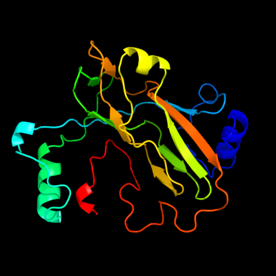
Phyre Home Retrieve Phyre Job Id
Emaill.a.kelley@imperial.ac.uk
DescriptionP28248
DateThu Jan 5 11:44:33 GMT 2012
Unique Job ID87f406cc73db9c4f

Summary 

Top model
Image coloured by rainbow N → C terminus
Model (left) based on template d1xs1a_
Top template information
Fold:beta-clip
Superfamily:dUTPase-like
Family:dUTPase-like
Confidence and coverage
Confidence:100.0% Coverage:100%
193 residues (100% of your sequence) have been modelled with 100.0% confidence by the single highest scoring template.
3D viewing
Interactive 3D view in Jmol

Sequence analysis 

Secondary structure and disorder prediction 

   1........10.........20.........30.........40.........50.........60
Sequence  MRLCDRDIEAWLDEGRLSINPRPPVERINGATVDVRLGNKFRTFRGHTAAFIDLSGPKDE
Secondary structure 


SS confidence 


Disorder  ????


?


??????
Disorder confidence 


   .........70.........80.........90.........100.........110.........120
Sequence  VSAALDRVMSDEIVLDEGEAFYLHPGELALAVTLESVTLPADLVGWLDGRSSLARLGLMV
Secondary structure 


SS confidence 


Disorder  ????


Disorder confidence 


   .........130.........140.........150.........160.........170.........180
Sequence  HVTAHRIDPGWSGCIVLEFYNSGKLPLALRPGMLIGALSFEPLSGPAVRPYNRREDAKYR
Secondary structure 


SS confidence 


Disorder 


???????????
Disorder confidence 


   .........190...
Sequence  NQQGAVASRIDKD
Secondary structure 


SS confidence 


Disorder  ?????
???????
Disorder confidence 


Confidence Key
High(9)                    Low (0)
?Disordered
Alpha helix
Beta strand

Domain analysis 

Hover over an aligned region to see model and summary info

Please note, only up to the top 20 hits are modelled to reduce computer load

RankAligned region

PDB 1xs1 chain A

3D model

Region: 1 - 193
Aligned: 193
Modelled: 193
Confidence: 100.0%
Identity: 100%
Fold: beta-clip
Superfamily: dUTPase-like
Family: dUTPase-like

Phyre2

PDB 2qxx chain A

3D model

Region: 1 - 193
Aligned: 184
Modelled: 193
Confidence: 100.0%
Identity: 44%
PDB header:hydrolase
Chain: A: PDB Molecule:deoxycytidine triphosphate deaminase;
PDBTitle: bifunctional dctp deaminase: dutpase from mycobacterium tuberculosis2 in complex with dttp

Phyre2

PDB 2qlp chain C

3D model

Region: 1 - 168
Aligned: 159
Modelled: 168
Confidence: 100.0%
Identity: 45%
PDB header:hydrolase
Chain: C: PDB Molecule:deoxycytidine triphosphate deaminase;
PDBTitle: bifunctional dctp deaminase:dutpase from mycobacterium tuberculosis,2 apo form

Phyre2

PDB 1pkh chain A

3D model

Region: 1 - 176
Aligned: 174
Modelled: 176
Confidence: 100.0%
Identity: 29%
Fold: beta-clip
Superfamily: dUTPase-like
Family: dUTPase-like

Phyre2

PDB 2r9q chain D

3D model

Region: 2 - 165
Aligned: 154
Modelled: 164
Confidence: 100.0%
Identity: 25%
PDB header:hydrolase
Chain: D: PDB Molecule:2'-deoxycytidine 5'-triphosphate deaminase;
PDBTitle: crystal structure of 2'-deoxycytidine 5'-triphosphate deaminase from2 agrobacterium tumefaciens

Phyre2

PDB 2yzj chain B

3D model

Region: 2 - 185
Aligned: 158
Modelled: 156
Confidence: 100.0%
Identity: 24%
PDB header:structural genomics, unknown function
Chain: B: PDB Molecule:167aa long hypothetical dutpase;
PDBTitle: crystal structure of dctp deaminase from sulfolobus tokodaii

Phyre2

PDB 3km3 chain B

3D model

Region: 2 - 161
Aligned: 146
Modelled: 149
Confidence: 100.0%
Identity: 29%
PDB header:hydrolase
Chain: B: PDB Molecule:deoxycytidine triphosphate deaminase;
PDBTitle: crystal structure of eoxycytidine triphosphate deaminase from2 anaplasma phagocytophilum at 2.1a resolution

Phyre2

PDB 3mbq chain C

3D model

Region: 29 - 179
Aligned: 115
Modelled: 115
Confidence: 99.9%
Identity: 26%
PDB header:hydrolase
Chain: C: PDB Molecule:deoxyuridine 5'-triphosphate nucleotidohydrolase;
PDBTitle: crystal structure of deoxyuridine 5-triphosphate nucleotidohydrolase2 from brucella melitensis, orthorhombic crystal form

Phyre2

PDB 1dun chain A

3D model

Region: 20 - 175
Aligned: 113
Modelled: 116
Confidence: 99.9%
Identity: 23%
Fold: beta-clip
Superfamily: dUTPase-like
Family: dUTPase-like

Phyre2

PDB 1f7r chain A

3D model

Region: 31 - 185
Aligned: 113
Modelled: 115
Confidence: 99.9%
Identity: 27%
Fold: beta-clip
Superfamily: dUTPase-like
Family: dUTPase-like

Phyre2

PDB 3ehw chain A

3D model

Region: 29 - 184
Aligned: 115
Modelled: 116
Confidence: 99.9%
Identity: 27%
PDB header:hydrolase
Chain: A: PDB Molecule:dutp pyrophosphatase;
PDBTitle: human dutpase in complex with alpha,beta-imido-dutp and mg2+:2 visualization of the full-length c-termini in all monomers and3 suggestion for an additional metal ion binding site

Phyre2

PDB 3tqz chain A

3D model

Region: 29 - 174
Aligned: 108
Modelled: 108
Confidence: 99.9%
Identity: 28%
PDB header:hydrolase
Chain: A: PDB Molecule:deoxyuridine 5'-triphosphate nucleotidohydrolase;
PDBTitle: structure of a deoxyuridine 5'-triphosphate nucleotidohydrolase (dut)2 from coxiella burnetii

Phyre2

PDB 1six chain A

3D model

Region: 29 - 184
Aligned: 115
Modelled: 116
Confidence: 99.9%
Identity: 31%
Fold: beta-clip
Superfamily: dUTPase-like
Family: dUTPase-like

Phyre2

PDB 1rnj chain A

3D model

Region: 29 - 175
Aligned: 109
Modelled: 109
Confidence: 99.9%
Identity: 21%
Fold: beta-clip
Superfamily: dUTPase-like
Family: dUTPase-like

Phyre2

PDB 2okd chain B

3D model

Region: 29 - 174
Aligned: 104
Modelled: 106
Confidence: 99.9%
Identity: 28%
PDB header:hydrolase
Chain: B: PDB Molecule:deoxyuridine 5'-triphosphate nucleotidohydrolase;
PDBTitle: high resolution crystal structures of vaccinia virus dutpase

Phyre2

PDB 3ca9 chain A

3D model

Region: 15 - 166
Aligned: 109
Modelled: 112
Confidence: 99.9%
Identity: 28%
PDB header:hydrolase
Chain: A: PDB Molecule:deoxyuridine triphosphatase;
PDBTitle: evolution of chlorella virus dutpase

Phyre2

PDB 1euw chain A

3D model

Region: 29 - 166
Aligned: 100
Modelled: 100
Confidence: 99.9%
Identity: 24%
Fold: beta-clip
Superfamily: dUTPase-like
Family: dUTPase-like

Phyre2

PDB 1sjn chain A

3D model

Region: 29 - 176
Aligned: 108
Modelled: 108
Confidence: 99.9%
Identity: 32%
Fold: beta-clip
Superfamily: dUTPase-like
Family: dUTPase-like

Phyre2

PDB 1f7d chain A

3D model

Region: 29 - 179
Aligned: 103
Modelled: 111
Confidence: 99.9%
Identity: 31%
Fold: beta-clip
Superfamily: dUTPase-like
Family: dUTPase-like

Phyre2

PDB 3c3i chain A

3D model

Region: 29 - 176
Aligned: 106
Modelled: 108
Confidence: 99.9%
Identity: 27%
PDB header:hydrolase
Chain: A: PDB Molecule:deoxyuridine triphosphatase;
PDBTitle: evolution of chlorella virus dutpase

Phyre2
1

d1xs1a_
2

c2qxxA_
3

c2qlpC_
4

d1pkha_
5

c2r9qD_
6

c2yzjB_
7

c3km3B_
8

c3mbqC_
9

d1duna_
10

d1f7ra_
11

c3ehwA_
12

c3tqzA_
13

d1sixa_
14

d1rnja_
15

c2okdB_
16

c3ca9A_
17

d1euwa_
18

d1sjna_
19

d1f7da_
20

c3c3iA_
21


22


23


24


25


26


27


28


29


30


31


32


33


34


35


36


37


38


39


40


41


42


43


44


45


46


47


48


49


50


Detailed template information 

#
Template Alignment Coverage3D Model Confidence
% i.d. Template Information
1d1xs1a_


100.0 100 Fold:beta-clip
Superfamily:dUTPase-like
Family:dUTPase-like
2c2qxxA_


100.0 44 PDB header:hydrolase
Chain: A: PDB Molecule:deoxycytidine triphosphate deaminase;
PDBTitle: bifunctional dctp deaminase: dutpase from mycobacterium tuberculosis2 in complex with dttp
3c2qlpC_


100.0 45 PDB header:hydrolase
Chain: C: PDB Molecule:deoxycytidine triphosphate deaminase;
PDBTitle: bifunctional dctp deaminase:dutpase from mycobacterium tuberculosis,2 apo form
4d1pkha_


100.0 29 Fold:beta-clip
Superfamily:dUTPase-like
Family:dUTPase-like
5c2r9qD_


100.0 25 PDB header:hydrolase
Chain: D: PDB Molecule:2'-deoxycytidine 5'-triphosphate deaminase;
PDBTitle: crystal structure of 2'-deoxycytidine 5'-triphosphate deaminase from2 agrobacterium tumefaciens
6c2yzjB_


100.0 24 PDB header:structural genomics, unknown function
Chain: B: PDB Molecule:167aa long hypothetical dutpase;
PDBTitle: crystal structure of dctp deaminase from sulfolobus tokodaii
7c3km3B_


100.0 29 PDB header:hydrolase
Chain: B: PDB Molecule:deoxycytidine triphosphate deaminase;
PDBTitle: crystal structure of eoxycytidine triphosphate deaminase from2 anaplasma phagocytophilum at 2.1a resolution
8c3mbqC_


99.9 26 PDB header:hydrolase
Chain: C: PDB Molecule:deoxyuridine 5'-triphosphate nucleotidohydrolase;
PDBTitle: crystal structure of deoxyuridine 5-triphosphate nucleotidohydrolase2 from brucella melitensis, orthorhombic crystal form
9d1duna_


99.9 23 Fold:beta-clip
Superfamily:dUTPase-like
Family:dUTPase-like
10d1f7ra_


99.9 27 Fold:beta-clip
Superfamily:dUTPase-like
Family:dUTPase-like
11c3ehwA_


99.9 27 PDB header:hydrolase
Chain: A: PDB Molecule:dutp pyrophosphatase;
PDBTitle: human dutpase in complex with alpha,beta-imido-dutp and mg2+:2 visualization of the full-length c-termini in all monomers and3 suggestion for an additional metal ion binding site
12c3tqzA_


99.9 28 PDB header:hydrolase
Chain: A: PDB Molecule:deoxyuridine 5'-triphosphate nucleotidohydrolase;
PDBTitle: structure of a deoxyuridine 5'-triphosphate nucleotidohydrolase (dut)2 from coxiella burnetii
13d1sixa_


99.9 31 Fold:beta-clip
Superfamily:dUTPase-like
Family:dUTPase-like
14d1rnja_


99.9 21 Fold:beta-clip
Superfamily:dUTPase-like
Family:dUTPase-like
15c2okdB_


99.9 28 PDB header:hydrolase
Chain: B: PDB Molecule:deoxyuridine 5'-triphosphate nucleotidohydrolase;
PDBTitle: high resolution crystal structures of vaccinia virus dutpase
16c3ca9A_


99.9 28 PDB header:hydrolase
Chain: A: PDB Molecule:deoxyuridine triphosphatase;
PDBTitle: evolution of chlorella virus dutpase
17d1euwa_


99.9 24 Fold:beta-clip
Superfamily:dUTPase-like
Family:dUTPase-like
18d1sjna_


99.9 32 Fold:beta-clip
Superfamily:dUTPase-like
Family:dUTPase-like
19d1f7da_


99.9 31 Fold:beta-clip
Superfamily:dUTPase-like
Family:dUTPase-like
20c3c3iA_


99.9 27 PDB header:hydrolase
Chain: A: PDB Molecule:deoxyuridine triphosphatase;
PDBTitle: evolution of chlorella virus dutpase
21c3f4fB_


not modelled 99.9 23 PDB header:hydrolase
Chain: B: PDB Molecule:deoxyuridine 5'-triphosphate nucleotidohydrolase;
PDBTitle: crystal structure of dut1p, a dutpase from saccharomyces cerevisiae
22d1q5uz_


not modelled 99.9 30 Fold:beta-clip
Superfamily:dUTPase-like
Family:dUTPase-like
23d3ehwa1


not modelled 99.9 29 Fold:beta-clip
Superfamily:dUTPase-like
Family:dUTPase-like
24d1vyqa1


not modelled 99.9 19 Fold:beta-clip
Superfamily:dUTPase-like
Family:dUTPase-like
25c3lqwA_


not modelled 99.9 32 PDB header:hydrolase
Chain: A: PDB Molecule:deoxyuridine 5'-triphosphate nucleotidohydrolase;
PDBTitle: crystal structure of deoxyuridine 5-triphosphate2 nucleotidohydrolase from entamoeba histolytica
26c2p9oB_


not modelled 99.9 27 PDB header:hydrolase
Chain: B: PDB Molecule:dutp pyrophosphatase-like protein;
PDBTitle: structure of dutpase from arabidopsis thaliana
27c2bazA_


not modelled 99.9 21 PDB header:unknown function
Chain: A: PDB Molecule:hypothetical protein bsu20020;
PDBTitle: structure of yoss, a putative dutpase from bacillus subtilis
28c3h6xA_


not modelled 99.9 24 PDB header:hydrolase
Chain: A: PDB Molecule:dutpase;
PDBTitle: crystal structure of dutpase from streptococcus mutans
29c2d4nA_


not modelled 99.9 27 PDB header:hydrolase
Chain: A: PDB Molecule:du;
PDBTitle: crystal structure of m-pmv dutpase complexed with dupnpp, substrate2 analogue
30c3ecyA_


not modelled 99.8 24 PDB header:hydrolase
Chain: A: PDB Molecule:cg4584-pa, isoform a (bcdna.ld08534);
PDBTitle: crystal structural analysis of drosophila melanogaster dutpase
31d2bsya2


not modelled 99.8 19 Fold:beta-clip
Superfamily:dUTPase-like
Family:dUTPase-like
32d2bsya1


not modelled 99.8 24 Fold:beta-clip
Superfamily:dUTPase-like
Family:dUTPase-like
33c2bt1A_


not modelled 99.8 20 PDB header:hydrolase
Chain: A: PDB Molecule:deoxyuridine 5'-triphosphate nucleotidohydrolase;
PDBTitle: epstein barr virus dutpase in complex with a,b-imino dutp
34d1tula_


not modelled 25.3 10 Fold:beta-clip
Superfamily:Tlp20, baculovirus telokin-like protein
Family:Tlp20, baculovirus telokin-like protein
35d1vioa2


not modelled 13.7 14 Fold:Alpha-L RNA-binding motif
Superfamily:Alpha-L RNA-binding motif
Family:Pseudouridine synthase RsuA N-terminal domain
36d1dm9a_


not modelled 12.4 11 Fold:Alpha-L RNA-binding motif
Superfamily:Alpha-L RNA-binding motif
Family:Heat shock protein 15 kD
37c1dm9A_


not modelled 12.4 11 PDB header:structural genomics
Chain: A: PDB Molecule:hypothetical 15.5 kd protein in mrca-pcka
PDBTitle: heat shock protein 15 kd
38d1vqop1


not modelled 11.3 21 Fold:Ribosomal protein L19 (L19e)
Superfamily:Ribosomal protein L19 (L19e)
Family:Ribosomal protein L19 (L19e)
39d1uwfa1


not modelled 11.1 19 Fold:Common fold of diphtheria toxin/transcription factors/cytochrome f
Superfamily:Bacterial adhesins
Family:Pilus subunits
40c4a1cO_


not modelled 8.6 11 PDB header:ribosome
Chain: O: PDB Molecule:rpl19;
PDBTitle: t.thermophila 60s ribosomal subunit in complex with2 initiation factor 6. this file contains 5s rrna,3 5.8s rrna and proteins of molecule 4.
41c3iz5T_


not modelled 8.3 15 PDB header:ribosome
Chain: T: PDB Molecule:60s ribosomal protein l19 (l19e);
PDBTitle: localization of the large subunit ribosomal proteins into a 5.5 a2 cryo-em map of triticum aestivum translating 80s ribosome
42d1v54b1


not modelled 7.7 27 Fold:Cupredoxin-like
Superfamily:Cupredoxins
Family:Periplasmic domain of cytochrome c oxidase subunit II
43d2visc_


not modelled 7.7 17 Fold:Viral protein domain
Superfamily:Viral protein domain
Family:Influenza hemagglutinin headpiece
44d2viua_


not modelled 7.2 17 Fold:Viral protein domain
Superfamily:Viral protein domain
Family:Influenza hemagglutinin headpiece
45d1mqma_


not modelled 7.1 18 Fold:Viral protein domain
Superfamily:Viral protein domain
Family:Influenza hemagglutinin headpiece
46c1ha0A_


not modelled 6.2 17 PDB header:viral protein
Chain: A: PDB Molecule:protein (hemagglutinin precursor);
PDBTitle: hemagglutinin precursor ha0
47c2ebbA_


not modelled 6.0 36 PDB header:lyase
Chain: A: PDB Molecule:pterin-4-alpha-carbinolamine dehydratase;
PDBTitle: crystal structure of pterin-4-alpha-carbinolamine2 dehydratase (pterin carbinolamine dehydratase) from3 geobacillus kaustophilus hta426
48d2odgc1


not modelled 5.5 25 Fold:LEM/SAP HeH motif
Superfamily:LEM domain
Family:LEM domain
49d1zx5a1


not modelled 5.4 13 Fold:Double-stranded beta-helix
Superfamily:RmlC-like cupins
Family:Type I phosphomannose isomerase
50d2phcb1


not modelled 5.3 27 Fold:Cyclophilin-like
Superfamily:Cyclophilin-like
Family:PH0987 C-terminal domain-like

Binding site prediction 

Due to computational demand, binding site predictions are not run for batch jobs

If you want to predict binding sites, please manually submit your model of choice to 3DLigandSite


Phyre is for academic use only

Please cite: Protein structure prediction on the web: a case study using the Phyre server
Kelley LA and Sternberg MJE. Nature Protocols 4, 363 - 371 (2009) [pdf] [Import into BibTeX]
 
If you use the binding site predictions from 3DLigandSite, please also cite:
3DLigandSite: predicting ligand-binding sites using similar structures.
Wass MN, Kelley LA and Sternberg MJ Nucleic Acids Research 38, W469-73 (2010) [PubMed]
 
© Structural Bioinformatics Group
Imperial College London
Lawrence Kelley, Benjamin Jefferys
Disclaimer
Terms and Conditions
Component software
Template detection: HHpred 1.51
Secondary structure prediction: Psi-pred 2.5
Disorder prediction: Disopred 2.4
Transmembrane prediction: Memsat_SVM
Multi-template modelling and ab initio: Poing 1.0