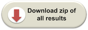
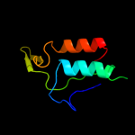
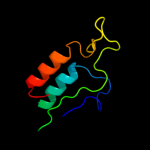
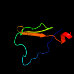
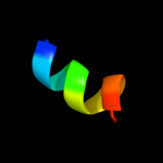
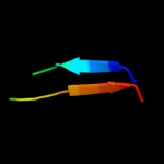
1 c2hjjA_
100.0
62
PDB header: structural genomics, unknown functionChain: A: PDB Molecule: hypothetical protein ykff;PDBTitle: solution nmr structure of protein ykff from escherichia coli.2 northeast structural genomics target er397.
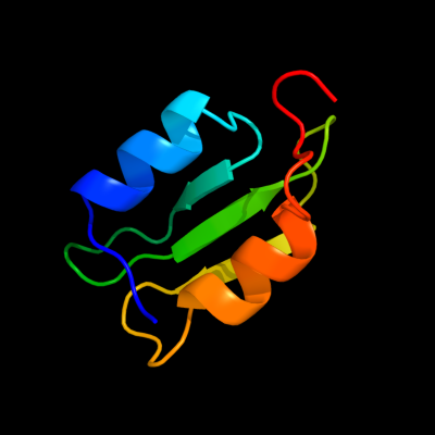
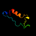
2 d2hjja1
100.0
62
Fold: dsRBD-likeSuperfamily: YcfA/nrd intein domainFamily: YkfF-like3 c3lrmB_
39.0
18
PDB header: hydrolaseChain: B: PDB Molecule: alpha-galactosidase 1;PDBTitle: structure of alfa-galactosidase from saccharomyces cerevisiae with2 raffinose
4 c3isyA_
32.9
33
PDB header: protein bindingChain: A: PDB Molecule: intracellular proteinase inhibitor;PDBTitle: crystal structure of an intracellular proteinase inhibitor (ipi,2 bsu11130) from bacillus subtilis at 2.61 a resolution
5 c3hx8A_
12.5
25
PDB header: isomeraseChain: A: PDB Molecule: putative ketosteroid isomerase;PDBTitle: crystal structure of putative ketosteroid isomerase2 (np_103587.1) from mesorhizobium loti at 1.45 a resolution
6 d1u2ca1
12.3
18
Fold: Immunoglobulin-like beta-sandwichSuperfamily: Cadherin-likeFamily: Dystroglycan, N-terminal domain7 d1uasa2
11.6
27
Fold: TIM beta/alpha-barrelSuperfamily: (Trans)glycosidasesFamily: Amylase, catalytic domain8 d1xdna_
11.5
13
Fold: ATP-graspSuperfamily: DNA ligase/mRNA capping enzyme, catalytic domainFamily: RNA ligase9 c1xs3A_
10.9
20
PDB header: structural genomics, unknown functionChain: A: PDB Molecule: hypothetical protein xc975;PDBTitle: solution structure analysis of the xc975 protein
10 c2dhmA_
9.9
12
PDB header: protein bindingChain: A: PDB Molecule: protein bola;PDBTitle: solution structure of the bola protein from escherichia coli
11 c3p1vB_
9.2
18
PDB header: hydrolaseChain: B: PDB Molecule: metallo-endopeptidase;PDBTitle: crystal structure of a metallo-endopeptidases (bacova_00663) from2 bacteroides ovatus at 1.93 a resolution
12 c1z64A_
8.5
57
PDB header: antimicrobial proteinChain: A: PDB Molecule: pleruocidin;PDBTitle: nmr solution structure of pleurocidin in dpc micelles
13 c2zt9F_
8.0
29
PDB header: photosynthesisChain: F: PDB Molecule: cytochrome b6-f complex subunit 7;PDBTitle: crystal structure of the cytochrome b6f complex from nostoc sp. pcc2 7120
14 c3tr3A_
7.6
20
PDB header: unknown functionChain: A: PDB Molecule: bola;PDBTitle: structure of a bola protein homologue from coxiella burnetii
15 c1yewF_
7.5
50
PDB header: oxidoreductase, membrane proteinChain: F: PDB Molecule: particulate methane monooxygenase, a subunit;PDBTitle: crystal structure of particulate methane monooxygenase
16 c3a23A_
7.4
31
PDB header: hydrolaseChain: A: PDB Molecule: putative secreted alpha-galactosidase;PDBTitle: crystal structure of beta-l-arabinopyranosidase complexed with d-2 galactose
17 c3ocjA_
7.3
26
PDB header: structural genomics, unknown functionChain: A: PDB Molecule: putative exported protein;PDBTitle: the crystal structure of a possilbe exported protein from bordetella2 parapertussis
18 c2kdnA_
7.1
13
PDB header: unknown functionChain: A: PDB Molecule: putative uncharacterized protein pfe0790c;PDBTitle: solution structure of pfe0790c, a putative bola-like2 protein from the protozoan parasite plasmodium falciparum.
19 c3o2eA_
6.7
13
PDB header: unknown functionChain: A: PDB Molecule: bola-like protein;PDBTitle: crystal structure of a bol-like protein from babesia bovis
20 d1x7fa2
6.5
31
Fold: TIM beta/alpha-barrelSuperfamily: (Trans)glycosidasesFamily: Outer surface protein, N-terminal domain21 c3ffeB_
not modelled
6.2
23
PDB header: biosynthetic proteinChain: B: PDB Molecule: acsd;PDBTitle: structure of achromobactin synthetase protein d, (acsd)
22 c2kz3A_
not modelled
6.1
22
PDB header: unknown functionChain: A: PDB Molecule: putative uncharacterized protein rad51l3;PDBTitle: backbone 1h, 13c, and 15n chemical shift assignments for human rad51d2 from 1 to 83
23 d1szna2
not modelled
5.6
27
Fold: TIM beta/alpha-barrelSuperfamily: (Trans)glycosidasesFamily: Amylase, catalytic domain24 c3gv2E_
not modelled
5.6
35
PDB header: viral proteinChain: E: PDB Molecule: fusion protein consisting of capsid protein p24,PDBTitle: x-ray structure of hexameric hiv-1 ca
25 c3chxF_
not modelled
5.3
46
PDB header: membrane proteinChain: F: PDB Molecule: pmoa;PDBTitle: crystal structure of methylosinus trichosporium ob3b2 particulate methane monooxygenase (pmmo)