| 1 |
|
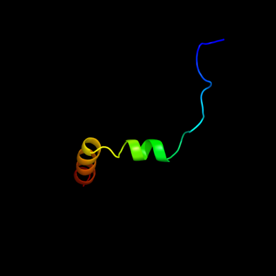
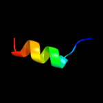
PDB 2voy chain B
Region: 100 - 137
Aligned: 38
Modelled: 38
Confidence: 18.6%
Identity: 8%
PDB header:hydrolase
Chain: B: PDB Molecule:sarcoplasmic/endoplasmic reticulum calcium
PDBTitle: cryoem model of copa, the copper transporting atpase from2 archaeoglobus fulgidus
Phyre2
| 2 |
|
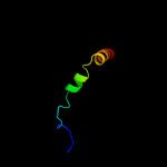
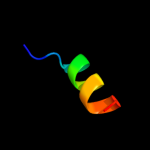
PDB 3eh4 chain A
Region: 68 - 136
Aligned: 69
Modelled: 69
Confidence: 11.9%
Identity: 9%
PDB header:oxidoreductase
Chain: A: PDB Molecule:cytochrome c oxidase subunit 1;
PDBTitle: structure of the reduced form of cytochrome ba3 oxidase from thermus2 thermophilus
Phyre2
| 3 |
|
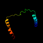
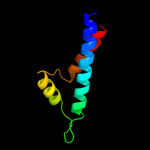
PDB 1xme chain A domain 1
Region: 68 - 136
Aligned: 69
Modelled: 69
Confidence: 9.2%
Identity: 9%
Fold: Cytochrome c oxidase subunit I-like
Superfamily: Cytochrome c oxidase subunit I-like
Family: Cytochrome c oxidase subunit I-like
Phyre2
| 4 |
|
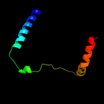
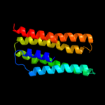
PDB 1q90 chain G
Region: 1 - 14
Aligned: 14
Modelled: 14
Confidence: 5.9%
Identity: 29%
Fold: Single transmembrane helix
Superfamily: PetG subunit of the cytochrome b6f complex
Family: PetG subunit of the cytochrome b6f complex
Phyre2
| 5 |
|
PDB 1q90 chain G
Region: 1 - 14
Aligned: 14
Modelled: 14
Confidence: 5.9%
Identity: 29%
PDB header:photosynthesis
Chain: G: PDB Molecule:cytochrome b6f complex subunit petg;
PDBTitle: structure of the cytochrome b6f (plastohydroquinone : plastocyanin2 oxidoreductase) from chlamydomonas reinhardtii
Phyre2
| 6 |
|
PDB 2d1k chain C
Region: 18 - 35
Aligned: 18
Modelled: 18
Confidence: 5.5%
Identity: 17%
PDB header:structural protein
Chain: C: PDB Molecule:metastasis suppressor protein 1;
PDBTitle: ternary complex of the wh2 domain of mim with actin-dnase i
Phyre2
| 7 |
|
PDB 1eys chain L
Region: 67 - 136
Aligned: 70
Modelled: 70
Confidence: 5.3%
Identity: 7%
Fold: Bacterial photosystem II reaction centre, L and M subunits
Superfamily: Bacterial photosystem II reaction centre, L and M subunits
Family: Bacterial photosystem II reaction centre, L and M subunits
Phyre2
| 8 |
|
PDB 1xio chain A
Region: 18 - 176
Aligned: 145
Modelled: 145
Confidence: 5.1%
Identity: 10%
Fold: Family A G protein-coupled receptor-like
Superfamily: Family A G protein-coupled receptor-like
Family: Bacteriorhodopsin-like
Phyre2
| 9 |
|
PDB 1xio chain A
Region: 18 - 176
Aligned: 145
Modelled: 145
Confidence: 5.1%
Identity: 10%
PDB header:signaling protein
Chain: A: PDB Molecule:anabaena sensory rhodopsin;
PDBTitle: anabaena sensory rhodopsin
Phyre2