Phyre Home Retrieve Phyre Job Id
Emaill.a.kelley@imperial.ac.uk
DescriptionP0ABC3
DateThu Jan 5 11:15:09 GMT 2012
Unique Job ID7caf33bffc53f426

Summary 

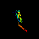
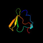
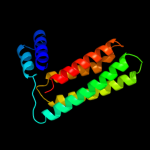
Top model
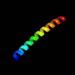
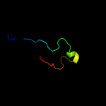
Image coloured by rainbow N → C terminus
Model (left) based on template c3bk6C_
Top template information
PDB header:membrane protein
Chain: C: PDB Molecule:ph stomatin;
PDBTitle: crystal structure of a core domain of stomatin from2 pyrococcus horikoshii
Confidence and coverage
Confidence: 99.9% Coverage: 47%
157 residues ( 47% of your sequence) have been modelled with 99.9% confidence by the single highest scoring template.
Additional confident templates have been detected (see Domain analysis) which cover other regions of your sequence.
264 residues ( 79%) could be modelled at >90% confidence using multiple-templates.
You may wish to try resubmitting your sequence in "intensive" mode to model more of your sequence.
3D viewing
Interactive 3D view in Jmol

Sequence analysis 

Secondary structure and disorder prediction 

   1........10.........20.........30.........40.........50.........60
Sequence  MRKSVIAIIIIVLVVLYMSVFVVKEGERGITLRFGKVLRDDDNKPLVYEPGLHFKIPFIE
Secondary structure 


SS confidence 


Disorder  ???


????


Disorder confidence 


   .........70.........80.........90.........100.........110.........120
Sequence  TVKMLDARIQTMDNQADRFVTKEKKDLIVDSYIKWRISDFSRYYLATGGGDISQAEVLLK
Secondary structure 


SS confidence 


Disorder 


?????

??


Disorder confidence 


   .........130.........140.........150.........160.........170.........180
Sequence  RKFSDRLRSEIGRLDVKDIVTDSRGRLTLEVRDALNSGSAGTEDEVTTPAADNAIAEAAE
Secondary structure 


SS confidence 


Disorder 


????????????????????
Disorder confidence 


   .........190.........200.........210.........220.........230.........240
Sequence  RVTAETKGKVPVINPNSMAALGIEVVDVRIKQINLPTEVSEAIYNRMRAEREAVARRHRS
Secondary structure 


SS confidence 


Disorder  ????????????????


??


Disorder confidence 


   .........250.........260.........270.........280.........290.........300
Sequence  QGQEEAEKLRATADYEVTRTLAEAERQGRIMRGEGDAEAAKLFADAFSKDPDFYAFIRSL
Secondary structure 
SS confidence 


Disorder 


?


?


?


Disorder confidence 


   .........310.........320.........330....
Sequence  RAYENSFSGNQDVMVMSPDSDFFRYMKTPTSATR
Secondary structure 


SS confidence 


Disorder 


?


?


????????
Disorder confidence 


Confidence Key
High(9)                    Low (0)
?Disordered
Alpha helix
Beta strand

Domain analysis 

Hover over an aligned region to see model and summary info

Please note, only up to the top 20 hits are modelled to reduce computer load

RankAligned region

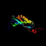
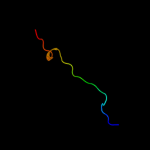
PDB 3bk6 chain C

3D model

Region: 58 - 259
Aligned: 157
Modelled: 163
Confidence: 99.9%
Identity: 25%
PDB header:membrane protein
Chain: C: PDB Molecule:ph stomatin;
PDBTitle: crystal structure of a core domain of stomatin from2 pyrococcus horikoshii

Phyre2

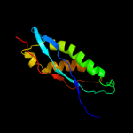
PDB 1win chain A

3D model

Region: 60 - 229
Aligned: 130
Modelled: 131
Confidence: 99.6%
Identity: 21%
Fold: EF-Ts domain-like
Superfamily: Band 7/SPFH domain
Family: Band 7/SPFH domain

Phyre2

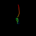
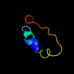
PDB 2rpb chain A

3D model

Region: 64 - 218
Aligned: 110
Modelled: 111
Confidence: 99.5%
Identity: 23%
PDB header:membrane protein
Chain: A: PDB Molecule:hypothetical membrane protein;
PDBTitle: the solution structure of membrane protein

Phyre2

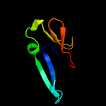
PDB 2zv4 chain O

3D model

Region: 21 - 289
Aligned: 260
Modelled: 267
Confidence: 98.8%
Identity: 15%
PDB header:structural protein
Chain: O: PDB Molecule:major vault protein;
PDBTitle: the structure of rat liver vault at 3.5 angstrom resolution

Phyre2

PDB 2kk7 chain A

3D model

Region: 254 - 290
Aligned: 37
Modelled: 37
Confidence: 38.9%
Identity: 24%
PDB header:hydrolase
Chain: A: PDB Molecule:v-type atp synthase subunit e;
PDBTitle: nmr solution structure of the n terminal domain of subunit e2 (e1-52) of a1ao atp synthase from methanocaldococcus3 jannaschii

Phyre2

PDB 3k5b chain B

3D model

Region: 226 - 292
Aligned: 66
Modelled: 67
Confidence: 33.7%
Identity: 24%
PDB header:hydrolase
Chain: B: PDB Molecule:v-type atp synthase, subunit (vapc-therm);
PDBTitle: crystal structure of the peripheral stalk of thermus thermophilus h+-2 atpase/synthase

Phyre2

PDB 3k5b chain E

3D model

Region: 223 - 309
Aligned: 79
Modelled: 82
Confidence: 16.7%
Identity: 13%
PDB header:hydrolase
Chain: E: PDB Molecule:v-type atp synthase subunit e;
PDBTitle: crystal structure of the peripheral stalk of thermus thermophilus h+-2 atpase/synthase

Phyre2

PDB 3sbt chain B

3D model

Region: 18 - 56
Aligned: 24
Modelled: 24
Confidence: 16.3%
Identity: 42%
PDB header:splicing
Chain: B: PDB Molecule:a1 cistron-splicing factor aar2;
PDBTitle: crystal structure of a aar2-prp8 complex

Phyre2

PDB 1js8 chain A domain 2

3D model

Region: 56 - 92
Aligned: 34
Modelled: 37
Confidence: 15.0%
Identity: 18%
Fold: C-terminal domain of mollusc hemocyanin
Superfamily: C-terminal domain of mollusc hemocyanin
Family: C-terminal domain of mollusc hemocyanin

Phyre2

PDB 2dii chain A

3D model

Region: 280 - 300
Aligned: 21
Modelled: 21
Confidence: 13.6%
Identity: 14%
PDB header:transcription
Chain: A: PDB Molecule:tfiih basal transcription factor complex p62
PDBTitle: solution structure of the bsd domain of human tfiih basal2 transcription factor complex p62 subunit

Phyre2

PDB 2ke4 chain A

3D model

Region: 254 - 293
Aligned: 40
Modelled: 40
Confidence: 12.2%
Identity: 20%
PDB header:membrane protein
Chain: A: PDB Molecule:cdc42-interacting protein 4;
PDBTitle: the nmr structure of the tc10 and cdc42 interacting domain2 of cip4

Phyre2

PDB 2dii chain A domain 1

3D model

Region: 280 - 300
Aligned: 21
Modelled: 21
Confidence: 10.9%
Identity: 14%
Fold: BSD domain-like
Superfamily: BSD domain-like
Family: BSD domain

Phyre2

PDB 3l4h chain A

3D model

Region: 34 - 84
Aligned: 51
Modelled: 51
Confidence: 9.8%
Identity: 10%
PDB header:protein binding
Chain: A: PDB Molecule:e3 ubiquitin-protein ligase hecw1;
PDBTitle: helical box domain and second ww domain of the human e3 ubiquitin-2 protein ligase hecw1

Phyre2

PDB 1lnl chain A domain 2

3D model

Region: 56 - 68
Aligned: 13
Modelled: 13
Confidence: 9.6%
Identity: 8%
Fold: C-terminal domain of mollusc hemocyanin
Superfamily: C-terminal domain of mollusc hemocyanin
Family: C-terminal domain of mollusc hemocyanin

Phyre2

PDB 1wd6 chain B

3D model

Region: 49 - 57
Aligned: 9
Modelled: 9
Confidence: 9.4%
Identity: 44%
PDB header:structural genomics, unknown function
Chain: B: PDB Molecule:protein ydhr;
PDBTitle: crystal structure of jw1657 from escherichia coli

Phyre2

PDB 1l2p chain A

3D model

Region: 227 - 289
Aligned: 56
Modelled: 63
Confidence: 8.8%
Identity: 20%
Fold: Single transmembrane helix
Superfamily: F1F0 ATP synthase subunit B, membrane domain
Family: F1F0 ATP synthase subunit B, membrane domain

Phyre2

PDB 1jey chain A domain 1

3D model

Region: 44 - 66
Aligned: 23
Modelled: 23
Confidence: 7.7%
Identity: 26%
Fold: SPOC domain-like
Superfamily: SPOC domain-like
Family: Ku70 subunit middle domain

Phyre2

PDB 1y4c chain A

3D model

Region: 145 - 289
Aligned: 142
Modelled: 142
Confidence: 7.2%
Identity: 11%
PDB header:de novo protein
Chain: A: PDB Molecule:maltose binding protein fused with designed
PDBTitle: designed helical protein fusion mbp

Phyre2

PDB 1nyo chain A

3D model

Region: 283 - 325
Aligned: 43
Modelled: 43
Confidence: 6.8%
Identity: 16%
Fold: FAS1 domain
Superfamily: FAS1 domain
Family: FAS1 domain

Phyre2

PDB 2ix0 chain A domain 3

3D model

Region: 47 - 99
Aligned: 52
Modelled: 53
Confidence: 6.7%
Identity: 6%
Fold: OB-fold
Superfamily: Nucleic acid-binding proteins
Family: Cold shock DNA-binding domain-like

Phyre2
1

c3bk6C_
2

d1wina_
3

c2rpbA_
4

c2zv4O_
5

c2kk7A_
6

c3k5bB_
7

c3k5bE_
8

c3sbtB_
9

d1js8a2
10

c2diiA_
11

c2ke4A_
12

d2diia1
13

c3l4hA_
14

d1lnla2
15

c1wd6B_
16

d1l2pa_
17

d1jeya1
18

c1y4cA_
19

d1nyoa_
20

d2ix0a3
21


22


23


24


25


26


27


28


29


30


31


32


33


34


35


36


37


38


39


40


41


42


43


Detailed template information 

#
Template Alignment Coverage3D Model Confidence
% i.d. Template Information
1c3bk6C_


99.9 25 PDB header:membrane protein
Chain: C: PDB Molecule:ph stomatin;
PDBTitle: crystal structure of a core domain of stomatin from2 pyrococcus horikoshii
2d1wina_


99.6 21 Fold:EF-Ts domain-like
Superfamily:Band 7/SPFH domain
Family:Band 7/SPFH domain
3c2rpbA_


99.5 23 PDB header:membrane protein
Chain: A: PDB Molecule:hypothetical membrane protein;
PDBTitle: the solution structure of membrane protein
4c2zv4O_


98.8 15 PDB header:structural protein
Chain: O: PDB Molecule:major vault protein;
PDBTitle: the structure of rat liver vault at 3.5 angstrom resolution
5c2kk7A_


38.9 24 PDB header:hydrolase
Chain: A: PDB Molecule:v-type atp synthase subunit e;
PDBTitle: nmr solution structure of the n terminal domain of subunit e2 (e1-52) of a1ao atp synthase from methanocaldococcus3 jannaschii
6c3k5bB_


33.7 24 PDB header:hydrolase
Chain: B: PDB Molecule:v-type atp synthase, subunit (vapc-therm);
PDBTitle: crystal structure of the peripheral stalk of thermus thermophilus h+-2 atpase/synthase
7c3k5bE_


16.7 13 PDB header:hydrolase
Chain: E: PDB Molecule:v-type atp synthase subunit e;
PDBTitle: crystal structure of the peripheral stalk of thermus thermophilus h+-2 atpase/synthase
8c3sbtB_


16.3 42 PDB header:splicing
Chain: B: PDB Molecule:a1 cistron-splicing factor aar2;
PDBTitle: crystal structure of a aar2-prp8 complex
9d1js8a2


15.0 18 Fold:C-terminal domain of mollusc hemocyanin
Superfamily:C-terminal domain of mollusc hemocyanin
Family:C-terminal domain of mollusc hemocyanin
10c2diiA_


13.6 14 PDB header:transcription
Chain: A: PDB Molecule:tfiih basal transcription factor complex p62
PDBTitle: solution structure of the bsd domain of human tfiih basal2 transcription factor complex p62 subunit
11c2ke4A_


12.2 20 PDB header:membrane protein
Chain: A: PDB Molecule:cdc42-interacting protein 4;
PDBTitle: the nmr structure of the tc10 and cdc42 interacting domain2 of cip4
12d2diia1


10.9 14 Fold:BSD domain-like
Superfamily:BSD domain-like
Family:BSD domain
13c3l4hA_


9.8 10 PDB header:protein binding
Chain: A: PDB Molecule:e3 ubiquitin-protein ligase hecw1;
PDBTitle: helical box domain and second ww domain of the human e3 ubiquitin-2 protein ligase hecw1
14d1lnla2


9.6 8 Fold:C-terminal domain of mollusc hemocyanin
Superfamily:C-terminal domain of mollusc hemocyanin
Family:C-terminal domain of mollusc hemocyanin
15c1wd6B_


9.4 44 PDB header:structural genomics, unknown function
Chain: B: PDB Molecule:protein ydhr;
PDBTitle: crystal structure of jw1657 from escherichia coli
16d1l2pa_


8.8 20 Fold:Single transmembrane helix
Superfamily:F1F0 ATP synthase subunit B, membrane domain
Family:F1F0 ATP synthase subunit B, membrane domain
17d1jeya1


7.7 26 Fold:SPOC domain-like
Superfamily:SPOC domain-like
Family:Ku70 subunit middle domain
18c1y4cA_


7.2 11 PDB header:de novo protein
Chain: A: PDB Molecule:maltose binding protein fused with designed
PDBTitle: designed helical protein fusion mbp
19d1nyoa_


6.8 16 Fold:FAS1 domain
Superfamily:FAS1 domain
Family:FAS1 domain
20d2ix0a3


6.7 6 Fold:OB-fold
Superfamily:Nucleic acid-binding proteins
Family:Cold shock DNA-binding domain-like
21c1jeqA_


not modelled 6.6 26 PDB header:dna binding protein
Chain: A: PDB Molecule:ku70;
PDBTitle: crystal structure of the ku heterodimer
22c2l9uA_


not modelled 6.6 25 PDB header:membrane protein
Chain: A: PDB Molecule:receptor tyrosine-protein kinase erbb-3;
PDBTitle: spatial structure of dimeric erbb3 transmembrane domain
23d1o70a2


not modelled 6.3 7 Fold:FAS1 domain
Superfamily:FAS1 domain
Family:FAS1 domain
24d1if1a_


not modelled 6.2 55 Fold:DNA/RNA-binding 3-helical bundle
Superfamily:"Winged helix" DNA-binding domain
Family:Interferon regulatory factor
25c2dllA_


not modelled 6.2 55 PDB header:cytokine
Chain: A: PDB Molecule:interferon regulatory factor 4;
PDBTitle: solution structure of the irf domain of human interferon2 regulator factors 4
26c3lj5H_


not modelled 6.0 12 PDB header:viral protein
Chain: H: PDB Molecule:portal protein;
PDBTitle: full length bacteriophage p22 portal protein
27c2ko6A_


not modelled 6.0 28 PDB header:structural genomics, unknown function
Chain: A: PDB Molecule:uncharacterized protein yihd;
PDBTitle: solution structure of protein sf3929 from shigella flexneri2 2a. northeast structural genomics consortium target3 sfr81/ontario center for structural proteomics target4 sf3929
28d1p5dx4


not modelled 5.9 15 Fold:TBP-like
Superfamily:Phosphoglucomutase, C-terminal domain
Family:Phosphoglucomutase, C-terminal domain
29d2ix0a1


not modelled 5.7 39 Fold:OB-fold
Superfamily:Nucleic acid-binding proteins
Family:Cold shock DNA-binding domain-like
30d2irfg_


not modelled 5.7 55 Fold:DNA/RNA-binding 3-helical bundle
Superfamily:"Winged helix" DNA-binding domain
Family:Interferon regulatory factor
31c2k88A_


not modelled 5.6 7 PDB header:hydrolase
Chain: A: PDB Molecule:vacuolar proton pump subunit g;
PDBTitle: association of subunit d (vma6p) and e (vma4p) with g2 (vma10p) and the nmr solution structure of subunit g (g1-3 59) of the saccharomyces cerevisiae v1vo atpase
32d1tf5a1


not modelled 5.5 39 Fold:Pre-protein crosslinking domain of SecA
Superfamily:Pre-protein crosslinking domain of SecA
Family:Pre-protein crosslinking domain of SecA
33d2fhza1


not modelled 5.5 30 Fold:ImmE5-like
Superfamily:ImmE5-like
Family:ImmE5-like
34d1ueba3


not modelled 5.5 27 Fold:OB-fold
Superfamily:Nucleic acid-binding proteins
Family:Cold shock DNA-binding domain-like
35d2f5tx2


not modelled 5.4 18 Fold:Phospholipase D/nuclease
Superfamily:Phospholipase D/nuclease
Family:TrmB middle domain-like
36d1gvia1


not modelled 5.4 21 Fold:Immunoglobulin-like beta-sandwich
Superfamily:E set domains
Family:E-set domains of sugar-utilizing enzymes
37d1tfra2


not modelled 5.3 22 Fold:PIN domain-like
Superfamily:PIN domain-like
Family:5' to 3' exonuclease catalytic domain
38c3bmaC_


not modelled 5.3 12 PDB header:ligase
Chain: C: PDB Molecule:d-alanyl-lipoteichoic acid synthetase;
PDBTitle: crystal structure of d-alanyl-lipoteichoic acid synthetase from2 streptococcus pneumoniae r6
39c2kseA_


not modelled 5.3 24 PDB header:transferase
Chain: A: PDB Molecule:sensor protein qsec;
PDBTitle: backbone structure of the membrane domain of e. coli2 histidine kinase receptor qsec, center for structures of3 membrane proteins (csmp) target 4311c
40c2iruA_


not modelled 5.2 31 PDB header:transferase
Chain: A: PDB Molecule:putative dna ligase-like protein rv0938/mt0965;
PDBTitle: crystal structure of the polymerase domain from mycobacterium2 tuberculosis ligase d
41d1h2ka_


not modelled 5.1 23 Fold:Double-stranded beta-helix
Superfamily:Clavaminate synthase-like
Family:Hypoxia-inducible factor HIF ihhibitor (FIH1)
42c1nohB_


not modelled 5.1 16 PDB header:viral protein
Chain: B: PDB Molecule:head morphogenesis protein;
PDBTitle: the structure of bacteriophage phi29 scaffolding protein2 gp7 after prohead assembly
43d2o4ta1


not modelled 5.1 12 Fold:Left-handed superhelix
Superfamily:BH3980-like
Family:BH3980-like

Binding site prediction 

Due to computational demand, binding site predictions are not run for batch jobs

If you want to predict binding sites, please manually submit your model of choice to 3DLigandSite


Phyre is for academic use only

Please cite: Protein structure prediction on the web: a case study using the Phyre server
Kelley LA and Sternberg MJE. Nature Protocols 4, 363 - 371 (2009) [pdf] [Import into BibTeX]
 
If you use the binding site predictions from 3DLigandSite, please also cite:
3DLigandSite: predicting ligand-binding sites using similar structures.
Wass MN, Kelley LA and Sternberg MJ Nucleic Acids Research 38, W469-73 (2010) [PubMed]
 
© Structural Bioinformatics Group
Imperial College London
Lawrence Kelley, Benjamin Jefferys
Disclaimer
Terms and Conditions
Component software
Template detection: HHpred 1.51
Secondary structure prediction: Psi-pred 2.5
Disorder prediction: Disopred 2.4
Transmembrane prediction: Memsat_SVM
Multi-template modelling and ab initio: Poing 1.0