| Secondary structure and disorder prediction | |
| | |
1 | . | . | . | . | . | . | . | . | 10 | . | . | . | . | . | . | . | . | . | 20 | . | . | . | . | . | . | . | . | . | 30 | . | . | . | . | . |
| Sequence | |
M | R | I | A | K | I | G | V | I | A | L | F | L | F | M | A | L | G | G | I | G | G | V | M | L | A | G | Y | T | F | I | L | R | A | G |
| Secondary structure | |
|  | | | | | | | | | | | | | | | | | | | | | | | | | | | | | | | |
|
|
| SS confidence | |
|
|
|
|
|
|
|
|
|
|
|
|
|
|
|
|
|
|
|
|
|
|
|
|
|
|
|
|
|
|
|
|
|
|
|
| Disorder | |
? | ? | ? | ? | ? | ? |
|
|
|
|
|
|
|
|
|
|
|
|
|
|
|
|
|
|
|
|
|
|
| ? | ? | ? | ? | ? | ? |
| Disorder confidence | |
|
|
|
|
|
|
|
|
|
|
|
|
|
|
|
|
|
|
|
|
|
|
|
|
|
|
|
|
|
|
|
|
|
|
|
| |
| Confidence Key |
| High(9) | |
|
|
|
|
|
|
|
|
|
Low (0) |
| ? | Disordered |
| Alpha helix |
  | Beta strand |
Hover over an aligned region to see model and summary info
Please note, only up to the top 20 hits are modelled to reduce computer load
|
| 1 |
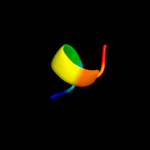
|
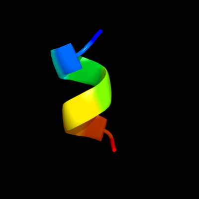
PDB 2lbg chain A
Region: 17 - 25
Aligned: 9
Modelled: 9
Confidence: 10.4%
Identity: 67%
PDB header:membrane protein
Chain: A: PDB Molecule:major prion protein;
PDBTitle: structure of the chr of the prion protein in dpc micelles
Phyre2
| 2 |
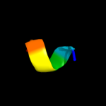
|
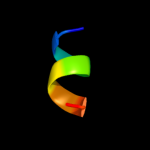
PDB 2axt chain J domain 1
Region: 3 - 21
Aligned: 19
Modelled: 19
Confidence: 9.4%
Identity: 32%
Fold: Single transmembrane helix
Superfamily: Photosystem II reaction center protein J, PsbJ
Family: PsbJ-like
Phyre2
| 3 |
|
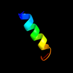
PDB 1eys chain H domain 2
Region: 1 - 16
Aligned: 16
Modelled: 16
Confidence: 8.1%
Identity: 31%
Fold: Single transmembrane helix
Superfamily: Photosystem II reaction centre subunit H, transmembrane region
Family: Photosystem II reaction centre subunit H, transmembrane region
Phyre2
| 4 |
|
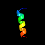
PDB 2kmc chain A
Region: 20 - 25
Aligned: 6
Modelled: 6
Confidence: 5.9%
Identity: 100%
PDB header:cell adhesion
Chain: A: PDB Molecule:fermitin family homolog 1;
PDBTitle: solution structure of the n-terminal domain of kindlin-1
Phyre2
| 5 |
|
PDB 2lgx chain A
Region: 20 - 25
Aligned: 6
Modelled: 6
Confidence: 5.3%
Identity: 100%
PDB header:cell adhesion
Chain: A: PDB Molecule:fermitin family homolog 2;
PDBTitle: nmr structure for kindle-2 n-terminus
Phyre2
|
| Detailed template information | |
Due to computational demand, binding site predictions are not run for batch jobs
If you want to predict binding sites, please manually submit your model of choice to 3DLigandSite
Phyre is for academic use only
| Please cite: Protein structure prediction on
the web: a case study using the Phyre server |
| Kelley LA and Sternberg MJE. Nature Protocols
4, 363 - 371 (2009) [pdf] [Import into BibTeX] |
| |
| If you use the binding site
predictions from 3DLigandSite, please also cite: |
| 3DLigandSite: predicting ligand-binding sites using similar structures. |
| Wass MN, Kelley LA and Sternberg
MJ Nucleic Acids Research 38, W469-73 (2010) [PubMed] |
| |
|
|
|
|