Phyre Home Retrieve Phyre Job Id
Emaill.a.kelley@imperial.ac.uk
DescriptionP64503
DateThu Jan 5 12:08:58 GMT 2012
Unique Job ID7453dbccab807e00

Summary 

Top model
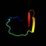
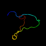
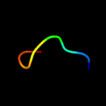
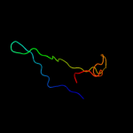
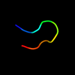
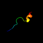
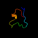
Image coloured by rainbow N → C terminus
Model (left) based on template d1pcia_
Top template information
Fold:Cysteine proteinases
Superfamily:Cysteine proteinases
Family:Papain-like
Confidence and coverage
Confidence: 23.3% Coverage: 37%
29 residues ( 37% of your sequence) have been modelled with 23.3% confidence by the single highest scoring template.
You may wish to submit your sequence to Phyrealarm. This will automatically scan your sequence every week for new potential templates as they appear in the Phyre2 library.
Please note: You must be registered and logged in to use Phyrealarm.
3D viewing
Interactive 3D view in Jmol

Sequence analysis 

Secondary structure and disorder prediction 

   1........10.........20.........30.........40.........50.........60
Sequence  MKTSVRIGAFEIDDGELHGESPGDRTLTIPCKSDPDLCMQLDAWDAETSIPALLNGEHSV
Secondary structure 


SS confidence 


Disorder  ???


???????


Disorder confidence 


   .........70........
Sequence  LYRTRYDQQSDAWIMRLA
Secondary structure 


SS confidence 


Disorder 


??


?
Disorder confidence 


Confidence Key
High(9)                    Low (0)
?Disordered
Alpha helix
Beta strand

Domain analysis 

Hover over an aligned region to see model and summary info

Please note, only up to the top 20 hits are modelled to reduce computer load

RankAligned region

PDB 1pci chain A

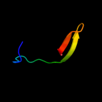
3D model

Region: 26 - 54
Aligned: 29
Modelled: 29
Confidence: 23.3%
Identity: 14%
Fold: Cysteine proteinases
Superfamily: Cysteine proteinases
Family: Papain-like

Phyre2

PDB 1xau chain A

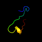
3D model

Region: 31 - 54
Aligned: 24
Modelled: 24
Confidence: 21.1%
Identity: 25%
Fold: Immunoglobulin-like beta-sandwich
Superfamily: Immunoglobulin
Family: I set domains

Phyre2

PDB 2hac chain A

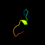
3D model

Region: 34 - 43
Aligned: 10
Modelled: 10
Confidence: 19.9%
Identity: 50%
PDB header:membrane protein
Chain: A: PDB Molecule:t-cell surface glycoprotein cd3 zeta chain;
PDBTitle: structure of zeta-zeta transmembrane dimer

Phyre2

PDB 3pnr chain B

3D model

Region: 19 - 54
Aligned: 36
Modelled: 36
Confidence: 16.4%
Identity: 19%
PDB header:hydrolase/hydrolase inhibitor
Chain: B: PDB Molecule:pbicp-c;
PDBTitle: structure of pbicp-c in complex with falcipain-2

Phyre2

PDB 1flt chain X

3D model

Region: 21 - 60
Aligned: 36
Modelled: 40
Confidence: 15.4%
Identity: 19%
Fold: Immunoglobulin-like beta-sandwich
Superfamily: Immunoglobulin
Family: I set domains

Phyre2

PDB 2aw2 chain A domain 1

3D model

Region: 31 - 54
Aligned: 24
Modelled: 24
Confidence: 15.1%
Identity: 33%
Fold: Immunoglobulin-like beta-sandwich
Superfamily: Immunoglobulin
Family: V set domains (antibody variable domain-like)

Phyre2

PDB 2c0y chain A

3D model

Region: 31 - 54
Aligned: 24
Modelled: 24
Confidence: 14.4%
Identity: 8%
PDB header:hydrolase
Chain: A: PDB Molecule:procathepsin s;
PDBTitle: the crystal structure of a cys25ala mutant of human2 procathepsin s

Phyre2

PDB 1ysj chain A domain 1

3D model

Region: 42 - 67
Aligned: 26
Modelled: 26
Confidence: 13.5%
Identity: 19%
Fold: Phosphorylase/hydrolase-like
Superfamily: Zn-dependent exopeptidases
Family: Bacterial dinuclear zinc exopeptidases

Phyre2

PDB 3raz chain A

3D model

Region: 41 - 62
Aligned: 22
Modelled: 22
Confidence: 12.9%
Identity: 18%
PDB header:oxidoreductase
Chain: A: PDB Molecule:thioredoxin-related protein;
PDBTitle: the crystal structure of thioredoxin-related protein from neisseria2 meningitidis serogroup b

Phyre2

PDB 3fju chain B

3D model

Region: 29 - 36
Aligned: 8
Modelled: 8
Confidence: 11.6%
Identity: 75%
PDB header:hydrolase/hydrolase inhibitor
Chain: B: PDB Molecule:carboxypeptidase a inhibitor;
PDBTitle: ascaris suum carboxypeptidase inhibitor in complex with human2 carboxypeptidase a1

Phyre2

PDB 2nn6 chain E domain 2

3D model

Region: 3 - 38
Aligned: 36
Modelled: 36
Confidence: 11.4%
Identity: 22%
Fold: Ribonuclease PH domain 2-like
Superfamily: Ribonuclease PH domain 2-like
Family: Ribonuclease PH domain 2-like

Phyre2

PDB 1xbs chain A

3D model

Region: 27 - 76
Aligned: 46
Modelled: 50
Confidence: 11.0%
Identity: 13%
PDB header:transcription, cell cycle
Chain: A: PDB Molecule:dim1-like protein;
PDBTitle: crystal structure of human dim2: a dim1-like protein

Phyre2

PDB 2opk chain C

3D model

Region: 66 - 77
Aligned: 12
Modelled: 9
Confidence: 10.0%
Identity: 33%
PDB header:isomerase
Chain: C: PDB Molecule:hypothetical protein;
PDBTitle: crystal structure of a putative mannose-6-phosphate isomerase2 (reut_a1446) from ralstonia eutropha jmp134 at 2.10 a resolution

Phyre2

PDB 2g8g chain A

3D model

Region: 4 - 48
Aligned: 45
Modelled: 45
Confidence: 9.9%
Identity: 20%
PDB header:virus
Chain: A: PDB Molecule:capsid;
PDBTitle: structurally mapping the diverse phenotype of adeno-2 associated virus serotype 4

Phyre2

PDB 1ppo chain A

3D model

Region: 30 - 54
Aligned: 25
Modelled: 25
Confidence: 8.7%
Identity: 16%
Fold: Cysteine proteinases
Superfamily: Cysteine proteinases
Family: Papain-like

Phyre2

PDB 2vsc chain D

3D model

Region: 25 - 72
Aligned: 40
Modelled: 48
Confidence: 7.8%
Identity: 15%
PDB header:cell adhesion
Chain: D: PDB Molecule:leukocyte surface antigen cd47;
PDBTitle: structure of the immunoglobulin-superfamily ectodomain of2 human cd47

Phyre2

PDB 1w4s chain A

3D model

Region: 28 - 72
Aligned: 45
Modelled: 45
Confidence: 6.9%
Identity: 13%
PDB header:nuclear protein
Chain: A: PDB Molecule:polybromo 1 protein;
PDBTitle: crystal structure of the proximal bah domain of polybromo

Phyre2

PDB 1b25 chain A domain 1

3D model

Region: 43 - 51
Aligned: 8
Modelled: 9
Confidence: 6.6%
Identity: 63%
Fold: Aldehyde ferredoxin oxidoreductase, C-terminal domains
Superfamily: Aldehyde ferredoxin oxidoreductase, C-terminal domains
Family: Aldehyde ferredoxin oxidoreductase, C-terminal domains

Phyre2

PDB 3iv6 chain C

3D model

Region: 1 - 18
Aligned: 18
Modelled: 18
Confidence: 6.4%
Identity: 28%
PDB header:oxidoreductase
Chain: C: PDB Molecule:putative zn-dependent alcohol dehydrogenase;
PDBTitle: crystal structure of putative zn-dependent alcohol dehydrogenases from2 rhodobacter sphaeroides.

Phyre2

PDB 2eq5 chain D

3D model

Region: 23 - 53
Aligned: 27
Modelled: 31
Confidence: 6.3%
Identity: 30%
PDB header:isomerase
Chain: D: PDB Molecule:228aa long hypothetical hydantoin racemase;
PDBTitle: crystal structure of hydantoin racemase from pyrococcus horikoshii ot3

Phyre2
1

d1pcia_
2

d1xaua_
3

c2hacA_
4

c3pnrB_
5

d1fltx_
6

d2aw2a1
7

c2c0yA_
8

d1ysja1
9

c3razA_
10

c3fjuB_
11

d2nn6e2
12

c1xbsA_
13

c2opkC_
14

c2g8gA_
15

d1ppoa_
16

c2vscD_
17

c1w4sA_
18

d1b25a1
19

c3iv6C_
20

c2eq5D_
21


22


23


24


25


26


27


28


Detailed template information 

#
Template Alignment Coverage3D Model Confidence
% i.d. Template Information
1d1pcia_


23.3 14 Fold:Cysteine proteinases
Superfamily:Cysteine proteinases
Family:Papain-like
2d1xaua_


21.1 25 Fold:Immunoglobulin-like beta-sandwich
Superfamily:Immunoglobulin
Family:I set domains
3c2hacA_


19.9 50 PDB header:membrane protein
Chain: A: PDB Molecule:t-cell surface glycoprotein cd3 zeta chain;
PDBTitle: structure of zeta-zeta transmembrane dimer
4c3pnrB_


16.4 19 PDB header:hydrolase/hydrolase inhibitor
Chain: B: PDB Molecule:pbicp-c;
PDBTitle: structure of pbicp-c in complex with falcipain-2
5d1fltx_


15.4 19 Fold:Immunoglobulin-like beta-sandwich
Superfamily:Immunoglobulin
Family:I set domains
6d2aw2a1


15.1 33 Fold:Immunoglobulin-like beta-sandwich
Superfamily:Immunoglobulin
Family:V set domains (antibody variable domain-like)
7c2c0yA_


14.4 8 PDB header:hydrolase
Chain: A: PDB Molecule:procathepsin s;
PDBTitle: the crystal structure of a cys25ala mutant of human2 procathepsin s
8d1ysja1


13.5 19 Fold:Phosphorylase/hydrolase-like
Superfamily:Zn-dependent exopeptidases
Family:Bacterial dinuclear zinc exopeptidases
9c3razA_


12.9 18 PDB header:oxidoreductase
Chain: A: PDB Molecule:thioredoxin-related protein;
PDBTitle: the crystal structure of thioredoxin-related protein from neisseria2 meningitidis serogroup b
10c3fjuB_


11.6 75 PDB header:hydrolase/hydrolase inhibitor
Chain: B: PDB Molecule:carboxypeptidase a inhibitor;
PDBTitle: ascaris suum carboxypeptidase inhibitor in complex with human2 carboxypeptidase a1
11d2nn6e2


11.4 22 Fold:Ribonuclease PH domain 2-like
Superfamily:Ribonuclease PH domain 2-like
Family:Ribonuclease PH domain 2-like
12c1xbsA_


11.0 13 PDB header:transcription, cell cycle
Chain: A: PDB Molecule:dim1-like protein;
PDBTitle: crystal structure of human dim2: a dim1-like protein
13c2opkC_


10.0 33 PDB header:isomerase
Chain: C: PDB Molecule:hypothetical protein;
PDBTitle: crystal structure of a putative mannose-6-phosphate isomerase2 (reut_a1446) from ralstonia eutropha jmp134 at 2.10 a resolution
14c2g8gA_


9.9 20 PDB header:virus
Chain: A: PDB Molecule:capsid;
PDBTitle: structurally mapping the diverse phenotype of adeno-2 associated virus serotype 4
15d1ppoa_


8.7 16 Fold:Cysteine proteinases
Superfamily:Cysteine proteinases
Family:Papain-like
16c2vscD_


7.8 15 PDB header:cell adhesion
Chain: D: PDB Molecule:leukocyte surface antigen cd47;
PDBTitle: structure of the immunoglobulin-superfamily ectodomain of2 human cd47
17c1w4sA_


6.9 13 PDB header:nuclear protein
Chain: A: PDB Molecule:polybromo 1 protein;
PDBTitle: crystal structure of the proximal bah domain of polybromo
18d1b25a1


6.6 63 Fold:Aldehyde ferredoxin oxidoreductase, C-terminal domains
Superfamily:Aldehyde ferredoxin oxidoreductase, C-terminal domains
Family:Aldehyde ferredoxin oxidoreductase, C-terminal domains
19c3iv6C_


6.4 28 PDB header:oxidoreductase
Chain: C: PDB Molecule:putative zn-dependent alcohol dehydrogenase;
PDBTitle: crystal structure of putative zn-dependent alcohol dehydrogenases from2 rhodobacter sphaeroides.
20c2eq5D_


6.3 30 PDB header:isomerase
Chain: D: PDB Molecule:228aa long hypothetical hydantoin racemase;
PDBTitle: crystal structure of hydantoin racemase from pyrococcus horikoshii ot3
21d2vzsa2


not modelled 6.3 12 Fold:Immunoglobulin-like beta-sandwich
Superfamily:beta-Galactosidase/glucuronidase domain
Family:beta-Galactosidase/glucuronidase domain
22c1ybiA_


not modelled 6.1 44 PDB header:toxin
Chain: A: PDB Molecule:non-toxin haemagglutinin ha34;
PDBTitle: crystal structure of ha33a, a neurotoxin-associated protein2 from clostridium botulinum type a
23d1oaza_


not modelled 5.9 23 Fold:Thioredoxin fold
Superfamily:Thioredoxin-like
Family:Thioltransferase
24d1a79a2


not modelled 5.8 31 Fold:MutS N-terminal domain-like
Superfamily:tRNA-intron endonuclease N-terminal domain-like
Family:tRNA-intron endonuclease N-terminal domain-like
25c2kllA_


not modelled 5.4 31 PDB header:cytokine
Chain: A: PDB Molecule:interleukin-33;
PDBTitle: solution structure of human interleukin-33
26d2tssa1


not modelled 5.4 29 Fold:OB-fold
Superfamily:Bacterial enterotoxins
Family:Superantigen toxins, N-terminal domain
27d1xmba1


not modelled 5.4 25 Fold:Phosphorylase/hydrolase-like
Superfamily:Zn-dependent exopeptidases
Family:Bacterial dinuclear zinc exopeptidases
28c2wmpB_


not modelled 5.3 33 PDB header:chaperone
Chain: B: PDB Molecule:papg protein;
PDBTitle: structure of the e. coli chaperone papd in complex with the pilin2 domain of the papgii adhesin

Binding site prediction 

Due to computational demand, binding site predictions are not run for batch jobs

If you want to predict binding sites, please manually submit your model of choice to 3DLigandSite


Phyre is for academic use only

Please cite: Protein structure prediction on the web: a case study using the Phyre server
Kelley LA and Sternberg MJE. Nature Protocols 4, 363 - 371 (2009) [pdf] [Import into BibTeX]
 
If you use the binding site predictions from 3DLigandSite, please also cite:
3DLigandSite: predicting ligand-binding sites using similar structures.
Wass MN, Kelley LA and Sternberg MJ Nucleic Acids Research 38, W469-73 (2010) [PubMed]
 
© Structural Bioinformatics Group
Imperial College London
Lawrence Kelley, Benjamin Jefferys
Disclaimer
Terms and Conditions
Component software
Template detection: HHpred 1.51
Secondary structure prediction: Psi-pred 2.5
Disorder prediction: Disopred 2.4
Transmembrane prediction: Memsat_SVM
Multi-template modelling and ab initio: Poing 1.0