| 1 |
|
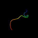
PDB 1y0k chain A domain 1
Region: 27 - 72
Aligned: 46
Modelled: 46
Confidence: 28.9%
Identity: 35%
Fold: Restriction endonuclease-like
Superfamily: Restriction endonuclease-like
Family: PA4535-like
Phyre2
| 2 |
|
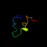
PDB 2dex chain X domain 1
Region: 42 - 53
Aligned: 12
Modelled: 12
Confidence: 14.9%
Identity: 50%
Fold: Common fold of diphtheria toxin/transcription factors/cytochrome f
Superfamily: Peptidylarginine deiminase Pad4, middle domain
Family: Peptidylarginine deiminase Pad4, middle domain
Phyre2
| 3 |
|
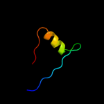
PDB 1nhp chain A domain 2
Region: 43 - 56
Aligned: 14
Modelled: 14
Confidence: 6.9%
Identity: 36%
Fold: FAD/NAD(P)-binding domain
Superfamily: FAD/NAD(P)-binding domain
Family: FAD/NAD-linked reductases, N-terminal and central domains
Phyre2
| 4 |
|
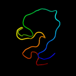
PDB 3nez chain B
Region: 22 - 54
Aligned: 30
Modelled: 33
Confidence: 5.8%
Identity: 33%
PDB header:fluorescent protein
Chain: B: PDB Molecule:mrojoa;
PDBTitle: mrojoa
Phyre2
| 5 |
|
PDB 2icr chain D
Region: 22 - 54
Aligned: 30
Modelled: 33
Confidence: 5.2%
Identity: 33%
PDB header:fluorescent protein
Chain: D: PDB Molecule:red fluorescent protein zoanrfp;
PDBTitle: red fluorescent protein zrfp574 from zoanthus sp.
Phyre2
| 6 |
|
PDB 1wqs chain A
Region: 1 - 40
Aligned: 40
Modelled: 40
Confidence: 5.0%
Identity: 25%
PDB header:hydrolase
Chain: A: PDB Molecule:3c-like protease;
PDBTitle: crystal structure of norovirus 3c-like protease
Phyre2
| 7 |
|
PDB 2vov chain A
Region: 4 - 35
Aligned: 32
Modelled: 32
Confidence: 5.0%
Identity: 34%
PDB header:metal-binding protein
Chain: A: PDB Molecule:surface-associated protein;
PDBTitle: an oxidized tryptophan facilitates copper-binding in2 methylococcus capsulatus secreted protein mope. the3 stucture of wild-type mope to 1.35aa
Phyre2