| Secondary structure and disorder prediction | |
| | |
1 | . | . | . | . | . | . | . | . | 10 | . | . | . | . | . | . | . | . | . | 20 | . | . | . | . | . | . | . | . | . | 30 | . | . | . | . | . | . | . | . | . | 40 | . | . | . | . | . | . | . | . | . | 50 | . | . | . | . | . | . | . | . | . | 60 |
| Sequence | |
M | C | P | E | C | F | F | L | M | L | F | F | C | G | Y | R | A | C | Y | C | S | S | S | F | S | S | S | S | S | S | S | S | S | S | S | F | R | S | S | P | A | Y | G | F | S | G | R | P | P | G | G | A | G | C | R | E | R | S | Q | R |
| Secondary structure | |
|
|  | | | | | | | | | | |
|
| | | | | |
|
|
|
|
|
|
| | | | | | | | | |
|
|
|
|
|
|
|
|
|
|
|
|
|
|
| | | | | | | | | |
| SS confidence | |
|
|
|
|
|
|
|
|
|
|
|
|
|
|
|
|
|
|
|
|
|
|
|
|
|
|
|
|
|
|
|
|
|
|
|
|
|
|
|
|
|
|
|
|
|
|
|
|
|
|
|
|
|
|
|
|
|
|
|
|
| Disorder | |
? | ? | ? |
|
|
|
|
|
|
|
|
|
|
|
|
|
|
|
|
|
| ? | ? | ? | ? | ? | ? | ? | ? | ? | ? | ? | ? | ? | ? | ? | ? | ? |
|
|
|
|
|
|
|
|
|
| ? | ? | ? | ? | ? | ? | ? | ? | ? | ? | ? |
|
| Disorder confidence | |
|
|
|
|
|
|
|
|
|
|
|
|
|
|
|
|
|
|
|
|
|
|
|
|
|
|
|
|
|
|
|
|
|
|
|
|
|
|
|
|
|
|
|
|
|
|
|
|
|
|
|
|
|
|
|
|
|
|
|
|
| |
| | |
. | . | . | . | . | . | . | . | . | 70 | . | . | . | . | . | . | . | . | . | 80 | . | . | . | . | . | . | . | . | . | 90 | . | . | . | . | . | . | . | . | . | 100 | . | . | . | . | . | . | . | . | . | 110 | . | . | . | . | . | . | . | . | . | 120 |
| Sequence | |
S | C | L | R | P | G | G | L | P | S | L | T | R | N | P | G | L | Q | R | P | F | R | S | R | R | L | C | R | A | V | A | C | A | P | G | I | P | A | K | G | R | R | D | V | R | G | N | A | V | S | Q | T | A | L | H | V | V | A | A | G |
| Secondary structure | |
| |
|
|
|
|
|
|
| | | | |
|
|
|
|
|
| | | | | | | | | | | | | | |
|
|
|
|
| | | | | | | | |
|
| | | | | | | | | | | |
|
| SS confidence | |
|
|
|
|
|
|
|
|
|
|
|
|
|
|
|
|
|
|
|
|
|
|
|
|
|
|
|
|
|
|
|
|
|
|
|
|
|
|
|
|
|
|
|
|
|
|
|
|
|
|
|
|
|
|
|
|
|
|
|
|
| Disorder | |
? | ? |
| ? |
|
|
|
|
|
| ? |
| ? |
|
| ? | ? | ? | ? | ? |
|
|
|
|
|
|
|
|
|
|
| ? | ? | ? | ? | ? | ? | ? | ? | ? | ? | ? |
| ? | ? | ? | ? | ? | ? |
| ? | ? | ? | ? |
| ? | ? | ? | ? |
|
| Disorder confidence | |
|
|
|
|
|
|
|
|
|
|
|
|
|
|
|
|
|
|
|
|
|
|
|
|
|
|
|
|
|
|
|
|
|
|
|
|
|
|
|
|
|
|
|
|
|
|
|
|
|
|
|
|
|
|
|
|
|
|
|
|
| |
| | |
. | . | . | . | . | . | . | . | . | 130 | . | . |
| Sequence | |
P | C | S | L | P | A | G | C | H | T | P | V |
| Secondary structure | |
|
|
|
|
|
|
|
|
|
|
|
|
| SS confidence | |
|
|
|
|
|
|
|
|
|
|
|
|
| Disorder | |
? |
|
|
|
| ? | ? | ? | ? | ? | ? | ? |
| Disorder confidence | |
|
|
|
|
|
|
|
|
|
|
|
|
| |
| Confidence Key |
| High(9) | |
|
|
|
|
|
|
|
|
|
Low (0) |
| ? | Disordered |
| Alpha helix |
  | Beta strand |
Hover over an aligned region to see model and summary info
Please note, only up to the top 20 hits are modelled to reduce computer load
|
| 1 |
|
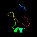
PDB 2l61 chain A
Region: 49 - 60
Aligned: 12
Modelled: 12
Confidence: 20.9%
Identity: 67%
PDB header:metal binding protein
Chain: A: PDB Molecule:ec protein i/ii;
PDBTitle: protein and metal cluster structure of the wheat metallothionein2 domain g-ec-1. the second part of the puzzle.
Phyre2
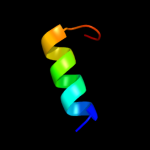
| 2 |
|
PDB 3ush chain B
Region: 15 - 54
Aligned: 40
Modelled: 40
Confidence: 8.1%
Identity: 28%
PDB header:structural genomics, unknown function
Chain: B: PDB Molecule:uncharacterized protein;
PDBTitle: crystal structure of the q2s0r5 protein from salinibacter ruber,2 northeast structural genomics consortium target srr207
Phyre2
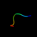
| 3 |
|
PDB 2l53 chain B
Region: 77 - 93
Aligned: 17
Modelled: 17
Confidence: 8.1%
Identity: 41%
PDB header:ca-binding protein/proton transport
Chain: B: PDB Molecule:voltage-gated sodium channel type v alpha isoform b
PDBTitle: solution nmr structure of apo-calmodulin in complex with the iq motif2 of human cardiac sodium channel nav1.5
Phyre2
| 4 |
|
PDB 1id3 chain B
Region: 40 - 46
Aligned: 7
Modelled: 7
Confidence: 6.5%
Identity: 57%
Fold: Histone-fold
Superfamily: Histone-fold
Family: Nucleosome core histones
Phyre2
|
| Detailed template information | |
Due to computational demand, binding site predictions are not run for batch jobs
If you want to predict binding sites, please manually submit your model of choice to 3DLigandSite
Phyre is for academic use only
| Please cite: Protein structure prediction on
the web: a case study using the Phyre server |
| Kelley LA and Sternberg MJE. Nature Protocols
4, 363 - 371 (2009) [pdf] [Import into BibTeX] |
| |
| If you use the binding site
predictions from 3DLigandSite, please also cite: |
| 3DLigandSite: predicting ligand-binding sites using similar structures. |
| Wass MN, Kelley LA and Sternberg
MJ Nucleic Acids Research 38, W469-73 (2010) [PubMed] |
| |
|
|
|
|