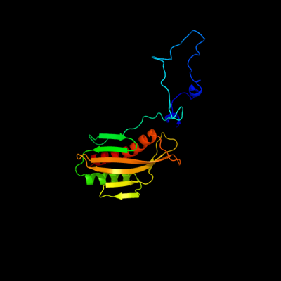
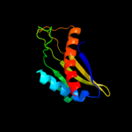
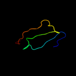
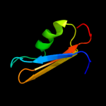
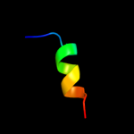
| 1 | c3tgoD_
|
|
 |
100.0 |
99 |
PDB header:membrane protein
Chain: D: PDB Molecule:lipoprotein 34;
PDBTitle: crystal structure of the e. coli bamcd complex
|
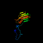
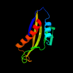
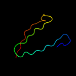
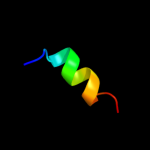
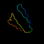
| 2 | c2yh5A_
|
|
 |
100.0 |
100 |
PDB header:lipid binding protein
Chain: A: PDB Molecule:dapx protein;
PDBTitle: structure of the c-terminal domain of bamc
|
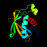
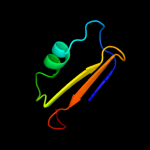
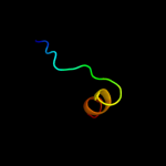
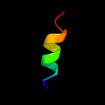
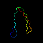
| 3 | c2lafA_
|
|
 |
99.9 |
100 |
PDB header:membrane protein
Chain: A: PDB Molecule:lipoprotein 34;
PDBTitle: nmr solution structure of the n-terminal domain of the e. coli2 lipoprotein bamc
|
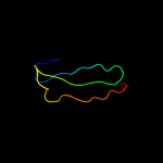
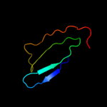
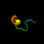
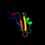
| 4 | c2yh6C_
|
|
 |
99.7 |
100 |
PDB header:lipid binding protein
Chain: C: PDB Molecule:lipoprotein 34;
PDBTitle: structure of the n-terminal domain of bamc from e. coli
|
| 5 | d1wi6a1
|
|
 |
62.2 |
20 |
Fold:Ferredoxin-like
Superfamily:RNA-binding domain, RBD
Family:Canonical RBD |
| 6 | d2d7na1
|
|
 |
50.3 |
16 |
Fold:Immunoglobulin-like beta-sandwich
Superfamily:E set domains
Family:Filamin repeat (rod domain) |
| 7 | d2e9ia1
|
|
 |
46.2 |
11 |
Fold:Immunoglobulin-like beta-sandwich
Superfamily:E set domains
Family:Filamin repeat (rod domain) |
| 8 | d1qfha1
|
|
 |
44.6 |
7 |
Fold:Immunoglobulin-like beta-sandwich
Superfamily:E set domains
Family:Filamin repeat (rod domain) |
| 9 | c3kdiA_
|
|
 |
40.0 |
20 |
PDB header:hormone receptor
Chain: A: PDB Molecule:putative uncharacterized protein at2g26040;
PDBTitle: structure of (+)-aba bound pyl2
|
| 10 | d2d7ma1
|
|
 |
38.1 |
12 |
Fold:Immunoglobulin-like beta-sandwich
Superfamily:E set domains
Family:Filamin repeat (rod domain) |
| 11 | d1wg4a_
|
|
 |
37.7 |
22 |
Fold:Ferredoxin-like
Superfamily:RNA-binding domain, RBD
Family:Canonical RBD |
| 12 | c3oquB_
|
|
 |
31.8 |
14 |
PDB header:hormone receptor
Chain: B: PDB Molecule:abscisic acid receptor pyl9;
PDBTitle: crystal structure of native abscisic acid receptor pyl9 with aba
|
| 13 | c3k90C_
|
|
 |
30.5 |
14 |
PDB header:hormone receptor, hydrolase regulator
Chain: C: PDB Molecule:putative uncharacterized protein;
PDBTitle: the abscisic acid receptor pyr1 in complex with abscisic acid
|
| 14 | c3p51A_
|
|
 |
30.3 |
16 |
PDB header:structural genomics, unknown function
Chain: A: PDB Molecule:uncharacterized protein;
PDBTitle: three-dimensional structure of protein q2y8n9_nitmu from nitrosospira2 multiformis, northeast structural genomics consortium target nmr118
|
| 15 | c3klxB_
|
|
 |
30.2 |
14 |
PDB header:hormone receptor
Chain: B: PDB Molecule:f3n23.20 protein;
PDBTitle: crystal structure of native abscisic acid receptor pyl3
|
| 16 | d2di8a1
|
|
 |
28.4 |
10 |
Fold:Immunoglobulin-like beta-sandwich
Superfamily:E set domains
Family:Filamin repeat (rod domain) |
| 17 | d2diba1
|
|
 |
28.4 |
20 |
Fold:Immunoglobulin-like beta-sandwich
Superfamily:E set domains
Family:Filamin repeat (rod domain) |
| 18 | d1wg1a_
|
|
 |
26.7 |
18 |
Fold:Ferredoxin-like
Superfamily:RNA-binding domain, RBD
Family:Canonical RBD |
| 19 | c1ksrA_
|
|
 |
26.3 |
13 |
PDB header:actin binding protein
Chain: A: PDB Molecule:gelation factor;
PDBTitle: the repeating segments of the f-actin cross-linking2 gelation factor (abp-120) have an immunoglobulin fold, nmr,3 20 structures
|
| 20 | c3iswA_
|
|
 |
24.7 |
13 |
PDB header:structural protein
Chain: A: PDB Molecule:filamin-a;
PDBTitle: crystal structure of filamin-a immunoglobulin-like repeat 21 bound to2 an n-terminal peptide of cftr
|
| 21 | c2brqB_ |
|
not modelled |
24.5 |
13 |
PDB header:structural protein
Chain: B: PDB Molecule:filamin a;
PDBTitle: crystal structure of the filamin a repeat 21 complexed with2 the integrin beta7 cytoplasmic tail peptide
|
| 22 | d2d7oa1 |
|
not modelled |
24.5 |
16 |
Fold:Immunoglobulin-like beta-sandwich
Superfamily:E set domains
Family:Filamin repeat (rod domain) |
| 23 | d2di9a1 |
|
not modelled |
23.5 |
16 |
Fold:Immunoglobulin-like beta-sandwich
Superfamily:E set domains
Family:Filamin repeat (rod domain) |
| 24 | c3e8vA_ |
|
not modelled |
22.6 |
20 |
PDB header:structural genomics, unknown function
Chain: A: PDB Molecule:possible transglutaminase-family protein;
PDBTitle: crystal structure of a possible transglutaminase-family2 protein proteolytic fragment from bacteroides fragilis
|
| 25 | c3qtjA_ |
|
not modelled |
22.2 |
25 |
PDB header:hormone receptor
Chain: A: PDB Molecule:abscisic acid receptor pyl10;
PDBTitle: crystal strcuture of aba receptor pyl10 (apo)
|
| 26 | c2le1A_ |
|
not modelled |
20.9 |
20 |
PDB header:structural genomics, unknown function
Chain: A: PDB Molecule:uncharacterized protein;
PDBTitle: solution nmr structure of tfu_2981 from thermobifida fusca, northeast2 structural genomics consortium target tfr85a
|
| 27 | c2pxgA_ |
|
not modelled |
20.6 |
17 |
PDB header:membrane protein
Chain: A: PDB Molecule:outer membrane protein;
PDBTitle: nmr solution structure of omla
|
| 28 | d1r89a3 |
|
not modelled |
20.4 |
19 |
Fold:Ferredoxin-like
Superfamily:PAP/Archaeal CCA-adding enzyme, C-terminal domain
Family:Archaeal tRNA CCA-adding enzyme |
| 29 | c2di7A_ |
|
not modelled |
20.1 |
22 |
PDB header:structural protein
Chain: A: PDB Molecule:bk158_1;
PDBTitle: solution structure of the filamin domain from human bk158_12 protein
|
| 30 | d2dmba1 |
|
not modelled |
20.0 |
7 |
Fold:Immunoglobulin-like beta-sandwich
Superfamily:E set domains
Family:Filamin repeat (rod domain) |
| 31 | c2ds4A_ |
|
not modelled |
19.7 |
9 |
PDB header:protein binding
Chain: A: PDB Molecule:tripartite motif protein 45;
PDBTitle: solution structure of the filamin domain from human2 tripartite motif protein 45
|
| 32 | d2d7pa1 |
|
not modelled |
19.4 |
11 |
Fold:Immunoglobulin-like beta-sandwich
Superfamily:E set domains
Family:Filamin repeat (rod domain) |
| 33 | c3hz7A_ |
|
not modelled |
19.2 |
18 |
PDB header:structural genomics, unknown function
Chain: A: PDB Molecule:uncharacterized protein;
PDBTitle: crystal structure of the sira-like protein (dsy4693) from2 desulfitobacterium hafniense, northeast structural genomics3 consortium target dhr2a
|
| 34 | d2cpda1 |
|
not modelled |
18.1 |
9 |
Fold:Ferredoxin-like
Superfamily:RNA-binding domain, RBD
Family:Canonical RBD |
| 35 | c2k9uA_ |
|
not modelled |
17.7 |
15 |
PDB header:structural protein
Chain: A: PDB Molecule:gamma filamin;
PDBTitle: solution nmr structure of the filamin-migfilin complex
|
| 36 | c2e44A_ |
|
not modelled |
17.7 |
15 |
PDB header:rna binding protein
Chain: A: PDB Molecule:insulin-like growth factor 2 mrna binding
PDBTitle: solution structure of rna binding domain in insulin-like2 growth factor 2 mrna binding protein 3
|
| 37 | d1uv7a_ |
|
not modelled |
17.5 |
10 |
Fold:RRF/tRNA synthetase additional domain-like
Superfamily:General secretion pathway protein M, EpsM
Family:General secretion pathway protein M, EpsM |
| 38 | c1uv7A_ |
|
not modelled |
17.5 |
10 |
PDB header:transport
Chain: A: PDB Molecule:general secretion pathway protein m;
PDBTitle: periplasmic domain of epsm from vibrio cholerae
|
| 39 | d3cnwa1 |
|
not modelled |
17.5 |
12 |
Fold:TBP-like
Superfamily:Bet v1-like
Family:Atu1531-like |
| 40 | d1wlha1 |
|
not modelled |
16.4 |
15 |
Fold:Immunoglobulin-like beta-sandwich
Superfamily:E set domains
Family:Filamin repeat (rod domain) |
| 41 | c2k7qA_ |
|
not modelled |
16.3 |
14 |
PDB header:structural protein
Chain: A: PDB Molecule:filamin-a;
PDBTitle: filamin a ig-like domains 18-19
|
| 42 | d2dj4a1 |
|
not modelled |
16.3 |
20 |
Fold:Immunoglobulin-like beta-sandwich
Superfamily:E set domains
Family:Filamin repeat (rod domain) |
| 43 | c2db8A_ |
|
not modelled |
16.3 |
13 |
PDB header:protein binding
Chain: A: PDB Molecule:tripartite motif protein 9, isoform 2;
PDBTitle: solution structures of the fn3 domain of human tripartite2 motif protein 9
|
| 44 | d2w0pa1 |
|
not modelled |
16.1 |
13 |
Fold:Immunoglobulin-like beta-sandwich
Superfamily:E set domains
Family:Filamin repeat (rod domain) |
| 45 | c2f7lA_ |
|
not modelled |
16.0 |
13 |
PDB header:isomerase
Chain: A: PDB Molecule:455aa long hypothetical phospho-sugar mutase;
PDBTitle: crystal structure of sulfolobus tokodaii2 phosphomannomutase/phosphoglucomutase
|
| 46 | c3c04A_ |
|
not modelled |
15.8 |
13 |
PDB header:isomerase
Chain: A: PDB Molecule:phosphomannomutase/phosphoglucomutase;
PDBTitle: structure of the p368g mutant of pmm/pgm from p. aeruginosa
|
| 47 | c3begB_ |
|
not modelled |
15.8 |
20 |
PDB header:transferase/splicing
Chain: B: PDB Molecule:splicing factor, arginine/serine-rich 1;
PDBTitle: crystal structure of sr protein kinase 1 complexed to its substrate2 asf/sf2
|
| 48 | c2npbA_ |
|
not modelled |
15.4 |
28 |
PDB header:oxidoreductase
Chain: A: PDB Molecule:selenoprotein w;
PDBTitle: nmr solution structure of mouse selw
|
| 49 | d2g7ja1 |
|
not modelled |
14.7 |
33 |
Fold:Secretion chaperone-like
Superfamily:YgaC/TfoX-N like
Family:YgaC-like |
| 50 | d1je3a_ |
|
not modelled |
13.7 |
16 |
Fold:IF3-like
Superfamily:SirA-like
Family:SirA-like |
| 51 | d2rera1 |
|
not modelled |
12.8 |
18 |
Fold:TBP-like
Superfamily:Bet v1-like
Family:oligoketide cyclase/dehydrase-like |
| 52 | d1axib2 |
|
not modelled |
12.6 |
18 |
Fold:Immunoglobulin-like beta-sandwich
Superfamily:Fibronectin type III
Family:Fibronectin type III |
| 53 | c2jxfA_ |
|
not modelled |
12.5 |
9 |
PDB header:viral protein, membrane protein
Chain: A: PDB Molecule:genome polyprotein;
PDBTitle: the solution structure of hcv ns4b(40-69)
|
| 54 | c3pdkB_ |
|
not modelled |
12.3 |
11 |
PDB header:isomerase
Chain: B: PDB Molecule:phosphoglucosamine mutase;
PDBTitle: crystal structure of phosphoglucosamine mutase from b. anthracis
|
| 55 | d1v05a_ |
|
not modelled |
12.1 |
18 |
Fold:Immunoglobulin-like beta-sandwich
Superfamily:E set domains
Family:Filamin repeat (rod domain) |
| 56 | c2ee6A_ |
|
not modelled |
12.1 |
15 |
PDB header:structural protein
Chain: A: PDB Molecule:filamin-b;
PDBTitle: solution structure of the 21th filamin domain from human2 filamin-b
|
| 57 | d2dica1 |
|
not modelled |
11.7 |
15 |
Fold:Immunoglobulin-like beta-sandwich
Superfamily:E set domains
Family:Filamin repeat (rod domain) |
| 58 | c1qfhB_ |
|
not modelled |
11.5 |
8 |
PDB header:actin binding protein
Chain: B: PDB Molecule:protein (gelation factor);
PDBTitle: dimerization of gelation factor from dictyostelium2 discoideum: crystal structure of rod domains 5 and 6
|
| 59 | d2d4ra1 |
|
not modelled |
11.3 |
5 |
Fold:TBP-like
Superfamily:Bet v1-like
Family:oligoketide cyclase/dehydrase-like |
| 60 | c2iyjA_ |
|
not modelled |
11.3 |
25 |
PDB header:isomerase
Chain: A: PDB Molecule:thiol disulfide interchange protein dsbc;
PDBTitle: crystal structure of the n-terminal dimer domain of e.coli2 dsbc
|
| 61 | d1x4xa1 |
|
not modelled |
11.2 |
15 |
Fold:Immunoglobulin-like beta-sandwich
Superfamily:Fibronectin type III
Family:Fibronectin type III |
| 62 | d1v5ja_ |
|
not modelled |
10.8 |
16 |
Fold:Immunoglobulin-like beta-sandwich
Superfamily:Fibronectin type III
Family:Fibronectin type III |
| 63 | d1iarb2 |
|
not modelled |
10.2 |
21 |
Fold:Immunoglobulin-like beta-sandwich
Superfamily:Fibronectin type III
Family:Fibronectin type III |
| 64 | c3rghA_ |
|
not modelled |
9.9 |
23 |
PDB header:cell adhesion
Chain: A: PDB Molecule:filamin-a;
PDBTitle: structure of filamin a immunoglobulin-like repeat 10 from homo sapiens
|
| 65 | d2diaa1 |
|
not modelled |
9.9 |
20 |
Fold:Immunoglobulin-like beta-sandwich
Superfamily:E set domains
Family:Filamin repeat (rod domain) |
| 66 | d2uubk1 |
|
not modelled |
9.9 |
16 |
Fold:Ribonuclease H-like motif
Superfamily:Translational machinery components
Family:Ribosomal protein L18 and S11 |
| 67 | d1qfha2 |
|
not modelled |
9.8 |
9 |
Fold:Immunoglobulin-like beta-sandwich
Superfamily:E set domains
Family:Filamin repeat (rod domain) |
| 68 | c3iswB_ |
|
not modelled |
9.8 |
13 |
PDB header:structural protein
Chain: B: PDB Molecule:filamin-a;
PDBTitle: crystal structure of filamin-a immunoglobulin-like repeat 21 bound to2 an n-terminal peptide of cftr
|
| 69 | c2w0pB_ |
|
not modelled |
9.8 |
13 |
PDB header:cell adhesion
Chain: B: PDB Molecule:filamin-a;
PDBTitle: crystal structure of the filamin a repeat 21 complexed with2 the migfilin peptide
|
| 70 | c2brqA_ |
|
not modelled |
9.8 |
13 |
PDB header:structural protein
Chain: A: PDB Molecule:filamin a;
PDBTitle: crystal structure of the filamin a repeat 21 complexed with2 the integrin beta7 cytoplasmic tail peptide
|
| 71 | c1n26A_ |
|
not modelled |
9.8 |
14 |
PDB header:cytokine
Chain: A: PDB Molecule:il-6 receptor alpha chain;
PDBTitle: crystal structure of the extra-cellular domains of human interleukin-62 receptor alpha chain
|
| 72 | d2dmca1 |
|
not modelled |
9.7 |
7 |
Fold:Immunoglobulin-like beta-sandwich
Superfamily:E set domains
Family:Filamin repeat (rod domain) |
| 73 | d2b79a1 |
|
not modelled |
9.7 |
24 |
Fold:TBP-like
Superfamily:Bet v1-like
Family:Smu440-like |
| 74 | c3cnkB_ |
|
not modelled |
9.6 |
9 |
PDB header:structural protein
Chain: B: PDB Molecule:filamin-a;
PDBTitle: crystal structure of the dimerization domain of human2 filamin a
|
| 75 | d1hn0a3 |
|
not modelled |
9.5 |
15 |
Fold:Hyaluronate lyase-like, C-terminal domain
Superfamily:Hyaluronate lyase-like, C-terminal domain
Family:Hyaluronate lyase-like, C-terminal domain |
| 76 | d1q38a_ |
|
not modelled |
9.1 |
24 |
Fold:Immunoglobulin-like beta-sandwich
Superfamily:Fibronectin type III
Family:Fibronectin type III |
| 77 | d1t1ea2 |
|
not modelled |
9.0 |
16 |
Fold:Ferredoxin-like
Superfamily:Protease propeptides/inhibitors
Family:Subtilase propeptides/inhibitors |
| 78 | d1q9ua_ |
|
not modelled |
9.0 |
27 |
Fold:TBP-like
Superfamily:TT1751-like
Family:TT1751-like |
| 79 | c3tfzB_ |
|
not modelled |
9.0 |
15 |
PDB header:biosynthetic protein
Chain: B: PDB Molecule:cyclase;
PDBTitle: crystal structure of zhui aromatase/cyclase from streptomcyes sp.2 r1128
|
| 80 | c2f3jA_ |
|
not modelled |
8.9 |
20 |
PDB header:transport protein
Chain: A: PDB Molecule:rna and export factor binding protein 2;
PDBTitle: the solution structure of the ref2-i mrna export factor2 (residues 1-155).
|
| 81 | c2glfB_ |
|
not modelled |
8.8 |
5 |
PDB header:hydrolase
Chain: B: PDB Molecule:probable m18-family aminopeptidase 1;
PDBTitle: crystal structure of aminipeptidase (m18 family) from thermotoga2 maritima
|
| 82 | d3begb1 |
|
not modelled |
8.8 |
20 |
Fold:Ferredoxin-like
Superfamily:RNA-binding domain, RBD
Family:Canonical RBD |
| 83 | c1wqaB_ |
|
not modelled |
8.8 |
21 |
PDB header:isomerase
Chain: B: PDB Molecule:phospho-sugar mutase;
PDBTitle: crystal structure of pyrococcus horikoshii2 phosphomannomutase/phosphoglucomutase complexed with mg2+
|
| 84 | d2qalk1 |
|
not modelled |
8.6 |
20 |
Fold:Ribonuclease H-like motif
Superfamily:Translational machinery components
Family:Ribosomal protein L18 and S11 |
| 85 | d1fyhb1 |
|
not modelled |
8.5 |
15 |
Fold:Immunoglobulin-like beta-sandwich
Superfamily:Fibronectin type III
Family:Fibronectin type III |
| 86 | d3d85d3 |
|
not modelled |
8.5 |
9 |
Fold:Immunoglobulin-like beta-sandwich
Superfamily:Fibronectin type III
Family:Fibronectin type III |
| 87 | c2rneA_ |
|
not modelled |
8.3 |
16 |
PDB header:rna binding protein
Chain: A: PDB Molecule:tia1 protein;
PDBTitle: solution structure of the second rna recognition motif2 (rrm) of tia-1
|
| 88 | d2q22a1 |
|
not modelled |
8.2 |
9 |
Fold:Ava3019-like
Superfamily:Ava3019-like
Family:Ava3019-like |
| 89 | d2bp3a1 |
|
not modelled |
7.9 |
13 |
Fold:Immunoglobulin-like beta-sandwich
Superfamily:E set domains
Family:Filamin repeat (rod domain) |
| 90 | c2j3sB_ |
|
not modelled |
7.9 |
14 |
PDB header:structural protein
Chain: B: PDB Molecule:filamin-a;
PDBTitle: crystal structure of the human filamin a ig domains 19 to2 21
|
| 91 | d1i7na1 |
|
not modelled |
7.8 |
21 |
Fold:PreATP-grasp domain
Superfamily:PreATP-grasp domain
Family:Synapsin domain |
| 92 | d2od4a1 |
|
not modelled |
7.8 |
20 |
Fold:Ferredoxin-like
Superfamily:Dimeric alpha+beta barrel
Family:Marine metagenome family DABB2 |
| 93 | c2dhxA_ |
|
not modelled |
7.6 |
24 |
PDB header:rna binding protein
Chain: A: PDB Molecule:poly (adp-ribose) polymerase family, member 10
PDBTitle: solution structure of the rrm domain in the human poly (adp-2 ribose) polymerase family, member 10 variant
|
| 94 | c2nqcA_ |
|
not modelled |
7.4 |
15 |
PDB header:immune system
Chain: A: PDB Molecule:filamin-c;
PDBTitle: crystal structure of ig-like domain 23 from human filamin c
|
| 95 | d2nqca1 |
|
not modelled |
7.4 |
15 |
Fold:Immunoglobulin-like beta-sandwich
Superfamily:E set domains
Family:Filamin repeat (rod domain) |
| 96 | c2yrzA_ |
|
not modelled |
7.2 |
19 |
PDB header:cell adhesion
Chain: A: PDB Molecule:integrin beta-4;
PDBTitle: solution structure of the fibronectin type iii domain of2 human integrin beta-4
|
| 97 | d1x5ia1 |
|
not modelled |
7.0 |
16 |
Fold:Immunoglobulin-like beta-sandwich
Superfamily:Fibronectin type III
Family:Fibronectin type III |
| 98 | d1a8ya3 |
|
not modelled |
6.9 |
7 |
Fold:Thioredoxin fold
Superfamily:Thioredoxin-like
Family:Calsequestrin |
| 99 | d2vkwa2 |
|
not modelled |
6.9 |
22 |
Fold:Immunoglobulin-like beta-sandwich
Superfamily:Fibronectin type III
Family:Fibronectin type III |