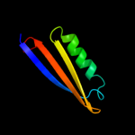
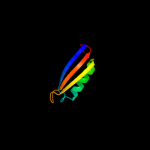
1 c3n7xA_
52.5
27
PDB header: virusChain: A: PDB Molecule: capsid protein;PDBTitle: crystal structure of penaeus stylirostris densovirus capsid
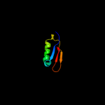
2 d1q5ya_
45.8
12
Fold: Ferredoxin-likeSuperfamily: ACT-likeFamily: Nickel responsive regulator NikR, C-terminal domain3 c2y3yC_
43.2
12
PDB header: transcriptionChain: C: PDB Molecule: putative nickel-responsive regulator;PDBTitle: holo-ni(ii) hpnikr is a symmetric tetramer containing four2 canonic square-planar ni(ii) ions at physiological ph
4 d2bj7a2
34.5
9
Fold: Ferredoxin-likeSuperfamily: ACT-likeFamily: Nickel responsive regulator NikR, C-terminal domain5 d1e8oa_
28.1
10
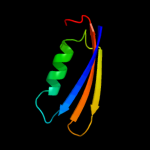
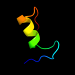
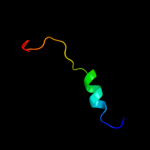
Fold: Signal recognition particle alu RNA binding heterodimer, SRP9/14Superfamily: Signal recognition particle alu RNA binding heterodimer, SRP9/14Family: Signal recognition particle alu RNA binding heterodimer, SRP9/146 d1914a1
22.7
11
Fold: Signal recognition particle alu RNA binding heterodimer, SRP9/14Superfamily: Signal recognition particle alu RNA binding heterodimer, SRP9/14Family: Signal recognition particle alu RNA binding heterodimer, SRP9/147 c2ca9B_
21.5
12
PDB header: transcriptional regulationChain: B: PDB Molecule: putative nickel-responsive regulator;PDBTitle: apo-nikr from helicobacter pylori in closed trans-2 conformation
8 c2bj3D_
20.9
11
PDB header: transcriptionChain: D: PDB Molecule: nickel responsive regulator;PDBTitle: nikr-apo
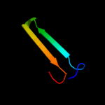
9 d2b5ib1
20.8
27
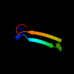
Fold: Immunoglobulin-like beta-sandwichSuperfamily: Fibronectin type IIIFamily: Fibronectin type III10 d3seba1
20.1
27
Fold: OB-foldSuperfamily: Bacterial enterotoxinsFamily: Superantigen toxins, N-terminal domain11 c3l4qA_
14.0
63
PDB header: viral protein/protein bindingChain: A: PDB Molecule: non-structural protein 1;PDBTitle: structural insights into phosphoinositide 3-kinase2 activation by the influenza a virus ns1 protein
12 c3d6rA_
13.7
63
PDB header: viral proteinChain: A: PDB Molecule: non-structural protein 1;PDBTitle: structure of an avian influenza a virus ns1 protein2 effector domain
13 d2gx9a1
13.6
63
Fold: Ns1 effector domain-likeSuperfamily: Ns1 effector domain-likeFamily: Ns1 effector domain-like14 d1c5cl2
12.7
16
Fold: Immunoglobulin-like beta-sandwichSuperfamily: ImmunoglobulinFamily: C1 set domains (antibody constant domain-like)15 c3f5tA_
11.3
63
PDB header: viral proteinChain: A: PDB Molecule: nonstructural protein 1;PDBTitle: x-ray structure of h5n1 ns1
16 c1914A_
10.6
11
PDB header: alu domainChain: A: PDB Molecule: signal recognition particle 9/14 fusion protein;PDBTitle: signal recognition particle alu rna binding heterodimer, srp9/14
17 d1bxta1
10.4
25
Fold: OB-foldSuperfamily: Bacterial enterotoxinsFamily: Superantigen toxins, N-terminal domain18 d1hyrc1
10.0
20
Fold: Immunoglobulin-like beta-sandwichSuperfamily: ImmunoglobulinFamily: C1 set domains (antibody constant domain-like)19 d1je6a1
8.5
17
Fold: Immunoglobulin-like beta-sandwichSuperfamily: ImmunoglobulinFamily: C1 set domains (antibody constant domain-like)20 c2e12B_
8.2
24
PDB header: translationChain: B: PDB Molecule: hypothetical protein xcc3642;PDBTitle: the crystal structure of xc5848 from xanthomonas campestris2 adopting a novel variant of sm-like motif
21 c2ns6A_
not modelled
7.4
19
PDB header: hydrolaseChain: A: PDB Molecule: mobilization protein a;PDBTitle: crystal structure of the minimal relaxase domain of moba2 from plasmid r1162
22 c3d33B_
not modelled
7.2
17
PDB header: unknown functionChain: B: PDB Molecule: domain of unknown function with an immunoglobulin-likePDBTitle: crystal structure of a duf3244 family protein with an immunoglobulin-2 like beta-sandwich fold (bvu_0276) from bacteroides vulgatus atcc3 8482 at 1.70 a resolution
23 c3mx7A_
not modelled
6.9
12
PDB header: apoptosisChain: A: PDB Molecule: fas apoptotic inhibitory molecule 1;PDBTitle: crystal structure analysis of human faim-ntd
24 d1tzoa_
not modelled
6.7
16
Fold: Anthrax protective antigenSuperfamily: Anthrax protective antigenFamily: Anthrax protective antigen25 d1irub_
not modelled
6.4
14
Fold: Ntn hydrolase-likeSuperfamily: N-terminal nucleophile aminohydrolases (Ntn hydrolases)Family: Proteasome subunits26 c1cqtJ_
not modelled
6.4
19
PDB header: gene regulation/dnaChain: J: PDB Molecule: pou domain, class 2, associating factor 1;PDBTitle: crystal structure of a ternary complex containing an oca-b2 peptide, the oct-1 pou domain, and an octamer element
27 c2p64B_
not modelled
6.1
16
PDB header: ligaseChain: B: PDB Molecule: f-box/wd repeat protein 1a;PDBTitle: d domain of b-trcp
28 d2q86b2
not modelled
6.1
13
Fold: Immunoglobulin-like beta-sandwichSuperfamily: ImmunoglobulinFamily: C1 set domains (antibody constant domain-like)29 c3sqgF_
not modelled
5.6
28
PDB header: transferaseChain: F: PDB Molecule: methyl-coenzyme m reductase, gamma subunit;PDBTitle: crystal structure of a methyl-coenzyme m reductase purified from black2 sea mats
30 c3n1uA_
not modelled
5.2
20
PDB header: hydrolaseChain: A: PDB Molecule: hydrolase, had superfamily, subfamily iii a;PDBTitle: structure of putative had superfamily (subfamily iii a) hydrolase from2 legionella pneumophila