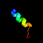
| 1 |
|
PDB 3mxn chain A
Region: 6 - 42
Aligned: 37
Modelled: 37
Confidence: 17.1%
Identity: 11%
PDB header:replication
Chain: A: PDB Molecule:recq-mediated genome instability protein 1;
PDBTitle: crystal structure of the rmi core complex
Phyre2
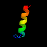
| 2 |
|
PDB 1bcg chain A
Region: 29 - 45
Aligned: 16
Modelled: 17
Confidence: 12.2%
Identity: 44%
Fold: Knottins (small inhibitors, toxins, lectins)
Superfamily: Scorpion toxin-like
Family: Long-chain scorpion toxins
Phyre2
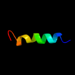
| 3 |
|
PDB 2l1w chain B
Region: 12 - 28
Aligned: 17
Modelled: 17
Confidence: 9.8%
Identity: 41%
PDB header:metal binding protein
Chain: B: PDB Molecule:vacuolar calcium atpase bca1 peptide;
PDBTitle: the solution structure of soybean calmodulin isoform 4 complexed with2 the vacuolar calcium atpase bca1 peptide
Phyre2
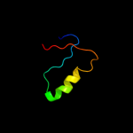
| 4 |
|
PDB 2rdc chain A
Region: 13 - 33
Aligned: 21
Modelled: 21
Confidence: 8.8%
Identity: 52%
PDB header:lipid binding protein
Chain: A: PDB Molecule:uncharacterized protein;
PDBTitle: crystal structure of a putative lipid binding protein (gsu0061) from2 geobacter sulfurreducens pca at 1.80 a resolution
Phyre2
| 5 |
|
PDB 3rnv chain A
Region: 23 - 43
Aligned: 21
Modelled: 21
Confidence: 8.1%
Identity: 33%
PDB header:hydrolase
Chain: A: PDB Molecule:helper component proteinase;
PDBTitle: structure of the autocatalytic cysteine protease domain of potyvirus2 helper-component proteinase
Phyre2
| 6 |
|
PDB 1okg chain A
Region: 2 - 40
Aligned: 39
Modelled: 38
Confidence: 6.7%
Identity: 21%
PDB header:transferase
Chain: A: PDB Molecule:possible 3-mercaptopyruvate sulfurtransferase;
PDBTitle: 3-mercaptopyruvate sulfurtransferase from leishmania major
Phyre2
| 7 |
|
PDB 2cl2 chain A
Region: 39 - 44
Aligned: 6
Modelled: 6
Confidence: 6.3%
Identity: 83%
PDB header:hydrolase
Chain: A: PDB Molecule:putative laminarinase;
PDBTitle: endo-1,3(4)-beta-glucanase from phanerochaete chrysosporium,2 solved using native sulfur sad, exhibiting intact3 heptasaccharide glycosylation
Phyre2