Phyre Home Retrieve Phyre Job Id
Emaill.a.kelley@imperial.ac.uk
DescriptionP0ACY9
DateThu Jan 5 11:19:27 GMT 2012
Unique Job ID4c5940d08162aa02

Summary 

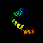
Top model
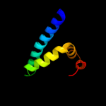
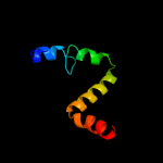
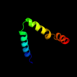
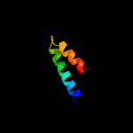
Image coloured by rainbow N → C terminus
Model (left) based on template c3ermD_
Top template information
PDB header:structural genomics, unknown function
Chain: D: PDB Molecule:uncharacterized conserved protein;
PDBTitle: the crystal structure of a conserved protein with unknown function2 from pseudomonas syringae pv. tomato str. dc3000
Confidence and coverage
Confidence: 99.9% Coverage: 68%
65 residues ( 68% of your sequence) have been modelled with 99.9% confidence by the single highest scoring template.
You may wish to submit your sequence to Phyrealarm. This will automatically scan your sequence every week for new potential templates as they appear in the Phyre2 library.
3D viewing
Interactive 3D view in Jmol

Sequence analysis 

Secondary structure and disorder prediction 

   1........10.........20.........30.........40.........50.........60
Sequence  MAVEVKYVVIREGEEKMSFTSKKEADAYDKMLDTADLLDTWLTNSPVQMEDEQREALSLW
Secondary structure 


SS confidence 


Disorder  ??


???


???????


Disorder confidence 


   .........70.........80.........90......
Sequence  LAEQKDVLSTILKTGKLPSPQVVGAESEEEDASHAA
Secondary structure 


SS confidence 


Disorder 


????????????????????????
Disorder confidence 


Confidence Key
High(9)                    Low (0)
?Disordered
Alpha helix
Beta strand

Domain analysis 

Hover over an aligned region to see model and summary info

Please note, only up to the top 20 hits are modelled to reduce computer load

RankAligned region

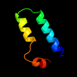
PDB 3erm chain D

3D model

Region: 21 - 85
Aligned: 65
Modelled: 65
Confidence: 99.9%
Identity: 23%
PDB header:structural genomics, unknown function
Chain: D: PDB Molecule:uncharacterized conserved protein;
PDBTitle: the crystal structure of a conserved protein with unknown function2 from pseudomonas syringae pv. tomato str. dc3000

Phyre2

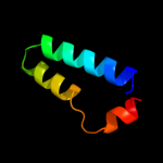
PDB 1vf6 chain C

3D model

Region: 30 - 73
Aligned: 44
Modelled: 44
Confidence: 52.6%
Identity: 20%
Fold: L27 domain
Superfamily: L27 domain
Family: L27 domain

Phyre2

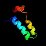
PDB 1vf6 chain C

3D model

Region: 30 - 73
Aligned: 44
Modelled: 44
Confidence: 52.6%
Identity: 20%
PDB header:protein binding/protein transport
Chain: C: PDB Molecule:maguk p55 subfamily member 5;
PDBTitle: 2.1 angstrom crystal structure of the pals-1-l27n and patj2 l27 heterodimer complex

Phyre2

PDB 1y76 chain B domain 1

3D model

Region: 30 - 73
Aligned: 44
Modelled: 44
Confidence: 51.2%
Identity: 23%
Fold: L27 domain
Superfamily: L27 domain
Family: L27 domain

Phyre2

PDB 1r45 chain A

3D model

Region: 18 - 76
Aligned: 47
Modelled: 58
Confidence: 45.5%
Identity: 15%
Fold: ADP-ribosylation
Superfamily: ADP-ribosylation
Family: ADP-ribosylating toxins

Phyre2

PDB 1puz chain A

3D model

Region: 31 - 81
Aligned: 51
Modelled: 51
Confidence: 40.8%
Identity: 12%
Fold: YgfY-like
Superfamily: YgfY-like
Family: YgfY-like

Phyre2

PDB 2jr5 chain A

3D model

Region: 31 - 85
Aligned: 55
Modelled: 55
Confidence: 33.9%
Identity: 9%
PDB header:structural genomics, unknown function
Chain: A: PDB Molecule:upf0350 protein vc_2471;
PDBTitle: solution structure of upf0350 protein vc_2471. northeast2 structural genomics target vcr36

Phyre2

PDB 1x6i chain B

3D model

Region: 31 - 85
Aligned: 55
Modelled: 55
Confidence: 32.5%
Identity: 11%
PDB header:structural genomics, unknown function
Chain: B: PDB Molecule:hypothetical protein ygfy;
PDBTitle: crystal structure of ygfy from escherichia coli

Phyre2

PDB 1fcd chain C domain 1

3D model

Region: 48 - 65
Aligned: 18
Modelled: 18
Confidence: 21.0%
Identity: 11%
Fold: Cytochrome c
Superfamily: Cytochrome c
Family: Two-domain cytochrome c

Phyre2

PDB 3bw8 chain B

3D model

Region: 19 - 76
Aligned: 46
Modelled: 53
Confidence: 18.7%
Identity: 20%
PDB header:transferase
Chain: B: PDB Molecule:mono-adp-ribosyltransferase c3;
PDBTitle: crystal structure of the clostridium limosum c3 exoenzyme

Phyre2

PDB 1f46 chain A

3D model

Region: 26 - 57
Aligned: 32
Modelled: 32
Confidence: 16.7%
Identity: 13%
Fold: TBP-like
Superfamily: Cell-division protein ZipA, C-terminal domain
Family: Cell-division protein ZipA, C-terminal domain

Phyre2

PDB 2kub chain A

3D model

Region: 29 - 65
Aligned: 37
Modelled: 37
Confidence: 16.5%
Identity: 24%
PDB header:structural protein
Chain: A: PDB Molecule:fimbriae-associated protein fap1;
PDBTitle: solution structure of the alpha subdomain of the major non-repeat unit2 of fap1 fimbriae of streptococcus parasanguis

Phyre2

PDB 2lm4 chain A

3D model

Region: 31 - 84
Aligned: 54
Modelled: 54
Confidence: 16.3%
Identity: 20%
PDB header:protein binding
Chain: A: PDB Molecule:succinate dehydrogenase assembly factor 2, mitochondrial;
PDBTitle: solution nmr structure of mitochondrial succinate dehydrogenase2 assembly factor 2 from saccharomyces cerevisiae, northeast structural3 genomics consortium target yt682a

Phyre2

PDB 3rgu chain A

3D model

Region: 29 - 65
Aligned: 37
Modelled: 37
Confidence: 16.3%
Identity: 24%
PDB header:structural protein
Chain: A: PDB Molecule:fimbriae-associated protein fap1;
PDBTitle: structure of fap-nra at ph 5.0

Phyre2

PDB 1fcd chain C domain 2

3D model

Region: 36 - 65
Aligned: 30
Modelled: 30
Confidence: 16.3%
Identity: 20%
Fold: Cytochrome c
Superfamily: Cytochrome c
Family: Two-domain cytochrome c

Phyre2

PDB 1kx7 chain A

3D model

Region: 48 - 65
Aligned: 18
Modelled: 18
Confidence: 16.1%
Identity: 22%
Fold: Cytochrome c
Superfamily: Cytochrome c
Family: monodomain cytochrome c

Phyre2

PDB 1wh7 chain A

3D model

Region: 16 - 38
Aligned: 23
Modelled: 23
Confidence: 14.8%
Identity: 35%
Fold: DNA/RNA-binding 3-helical bundle
Superfamily: Homeodomain-like
Family: Homeodomain

Phyre2

PDB 1ojq chain A

3D model

Region: 19 - 84
Aligned: 54
Modelled: 66
Confidence: 14.7%
Identity: 15%
Fold: ADP-ribosylation
Superfamily: ADP-ribosylation
Family: ADP-ribosylating toxins

Phyre2

PDB 1c75 chain A

3D model

Region: 49 - 65
Aligned: 17
Modelled: 17
Confidence: 14.0%
Identity: 41%
Fold: Cytochrome c
Superfamily: Cytochrome c
Family: monodomain cytochrome c

Phyre2

PDB 3cs5 chain B

3D model

Region: 43 - 74
Aligned: 32
Modelled: 32
Confidence: 13.4%
Identity: 22%
PDB header:photosynthesis
Chain: B: PDB Molecule:phycobilisome degradation protein nbla;
PDBTitle: nbla protein from synechococcus elongatus pcc 7942

Phyre2
1

c3ermD_
2

d1vf6c_
3

c1vf6C_
4

d1y76b1
5

d1r45a_
6

d1puza_
7

c2jr5A_
8

c1x6iB_
9

d1fcdc1
10

c3bw8B_
11

d1f46a_
12

c2kubA_
13

c2lm4A_
14

c3rguA_
15

d1fcdc2
16

d1kx7a_
17

d1wh7a_
18

d1ojqa_
19

d1c75a_
20

c3cs5B_
21


22


23


24


25


26


27


28


29


30


31


32


33


34


35


Detailed template information 

#
Template Alignment Coverage3D Model Confidence
% i.d. Template Information
1c3ermD_


99.9 23 PDB header:structural genomics, unknown function
Chain: D: PDB Molecule:uncharacterized conserved protein;
PDBTitle: the crystal structure of a conserved protein with unknown function2 from pseudomonas syringae pv. tomato str. dc3000
2d1vf6c_


52.6 20 Fold:L27 domain
Superfamily:L27 domain
Family:L27 domain
3c1vf6C_


52.6 20 PDB header:protein binding/protein transport
Chain: C: PDB Molecule:maguk p55 subfamily member 5;
PDBTitle: 2.1 angstrom crystal structure of the pals-1-l27n and patj2 l27 heterodimer complex
4d1y76b1


51.2 23 Fold:L27 domain
Superfamily:L27 domain
Family:L27 domain
5d1r45a_


45.5 15 Fold:ADP-ribosylation
Superfamily:ADP-ribosylation
Family:ADP-ribosylating toxins
6d1puza_


40.8 12 Fold:YgfY-like
Superfamily:YgfY-like
Family:YgfY-like
7c2jr5A_


33.9 9 PDB header:structural genomics, unknown function
Chain: A: PDB Molecule:upf0350 protein vc_2471;
PDBTitle: solution structure of upf0350 protein vc_2471. northeast2 structural genomics target vcr36
8c1x6iB_


32.5 11 PDB header:structural genomics, unknown function
Chain: B: PDB Molecule:hypothetical protein ygfy;
PDBTitle: crystal structure of ygfy from escherichia coli
9d1fcdc1


21.0 11 Fold:Cytochrome c
Superfamily:Cytochrome c
Family:Two-domain cytochrome c
10c3bw8B_


18.7 20 PDB header:transferase
Chain: B: PDB Molecule:mono-adp-ribosyltransferase c3;
PDBTitle: crystal structure of the clostridium limosum c3 exoenzyme
11d1f46a_


16.7 13 Fold:TBP-like
Superfamily:Cell-division protein ZipA, C-terminal domain
Family:Cell-division protein ZipA, C-terminal domain
12c2kubA_


16.5 24 PDB header:structural protein
Chain: A: PDB Molecule:fimbriae-associated protein fap1;
PDBTitle: solution structure of the alpha subdomain of the major non-repeat unit2 of fap1 fimbriae of streptococcus parasanguis
13c2lm4A_


16.3 20 PDB header:protein binding
Chain: A: PDB Molecule:succinate dehydrogenase assembly factor 2, mitochondrial;
PDBTitle: solution nmr structure of mitochondrial succinate dehydrogenase2 assembly factor 2 from saccharomyces cerevisiae, northeast structural3 genomics consortium target yt682a
14c3rguA_


16.3 24 PDB header:structural protein
Chain: A: PDB Molecule:fimbriae-associated protein fap1;
PDBTitle: structure of fap-nra at ph 5.0
15d1fcdc2


16.3 20 Fold:Cytochrome c
Superfamily:Cytochrome c
Family:Two-domain cytochrome c
16d1kx7a_


16.1 22 Fold:Cytochrome c
Superfamily:Cytochrome c
Family:monodomain cytochrome c
17d1wh7a_


14.8 35 Fold:DNA/RNA-binding 3-helical bundle
Superfamily:Homeodomain-like
Family:Homeodomain
18d1ojqa_


14.7 15 Fold:ADP-ribosylation
Superfamily:ADP-ribosylation
Family:ADP-ribosylating toxins
19d1c75a_


14.0 41 Fold:Cytochrome c
Superfamily:Cytochrome c
Family:monodomain cytochrome c
20c3cs5B_


13.4 22 PDB header:photosynthesis
Chain: B: PDB Molecule:phycobilisome degradation protein nbla;
PDBTitle: nbla protein from synechococcus elongatus pcc 7942
21d1vima_


not modelled 12.2 8 Fold:SIS domain
Superfamily:SIS domain
Family:mono-SIS domain
22d1cora_


not modelled 11.5 28 Fold:Cytochrome c
Superfamily:Cytochrome c
Family:monodomain cytochrome c
23c2bovB_


not modelled 11.3 17 PDB header:transferase
Chain: B: PDB Molecule:mono-adp-ribosyltransferase c3;
PDBTitle: molecular recognition of an adp-ribosylating clostridium2 botulinum c3 exoenzyme by rala gtpase
24d2c8aa1


not modelled 11.0 17 Fold:ADP-ribosylation
Superfamily:ADP-ribosylation
Family:ADP-ribosylating toxins
25d1lqta1


not modelled 9.9 24 Fold:FAD/NAD(P)-binding domain
Superfamily:FAD/NAD(P)-binding domain
Family:C-terminal domain of adrenodoxin reductase-like
26d1h1oa1


not modelled 8.3 22 Fold:Cytochrome c
Superfamily:Cytochrome c
Family:Two-domain cytochrome c
27c1u6hB_


not modelled 8.3 40 PDB header:cell adhesion
Chain: B: PDB Molecule:talin;
PDBTitle: vinculin head (0-258) in complex with the talin vinculin2 binding site 2 (849-879)
28c1qs2A_


not modelled 7.9 26 PDB header:toxin
Chain: A: PDB Molecule:adp-ribosyltransferase;
PDBTitle: crystal structure of vip2 with nad
29d1urfa_


not modelled 6.6 22 Fold:Long alpha-hairpin
Superfamily:HR1 repeat
Family:HR1 repeat
30c2c9eA_


not modelled 6.2 50 PDB header:photosynthesis
Chain: A: PDB Molecule:peridinin-chlorophyll a protein;
PDBTitle: peridinin-chlorophyll a protein, high-salt form
31d351ca_


not modelled 6.0 28 Fold:Cytochrome c
Superfamily:Cytochrome c
Family:monodomain cytochrome c
32c2d0sA_


not modelled 5.4 22 PDB header:electron transport
Chain: A: PDB Molecule:cytochrome c;
PDBTitle: crystal structure of the cytochrome c552 from moderate2 thermophilic bacterium, hydrogenophilus thermoluteolus
33c2kr1A_


not modelled 5.4 10 PDB header:ligase
Chain: A: PDB Molecule:ubiquitin protein ligase e3a;
PDBTitle: solution nmr structure of zinc binding n-terminal domain of ubiquitin-2 protein ligase e3a from homo sapiens. northeast structural genomics3 consortium (nesg) target hr3662
34c1skoA_


not modelled 5.2 23 PDB header:signaling protein
Chain: A: PDB Molecule:mitogen-activated protein kinase kinase 1
PDBTitle: mp1-p14 complex
35c1qs1D_


not modelled 5.1 24 PDB header:toxin
Chain: D: PDB Molecule:adp-ribosyltransferase;
PDBTitle: crystal structure of vegetative insecticidal protein2 (vip2)

Binding site prediction 

Due to computational demand, binding site predictions are not run for batch jobs

If you want to predict binding sites, please manually submit your model of choice to 3DLigandSite


Phyre is for academic use only

Please cite: Protein structure prediction on the web: a case study using the Phyre server
Kelley LA and Sternberg MJE. Nature Protocols 4, 363 - 371 (2009) [pdf] [Import into BibTeX]
 
If you use the binding site predictions from 3DLigandSite, please also cite:
3DLigandSite: predicting ligand-binding sites using similar structures.
Wass MN, Kelley LA and Sternberg MJ Nucleic Acids Research 38, W469-73 (2010) [PubMed]
 
© Structural Bioinformatics Group
Imperial College London
Lawrence Kelley, Benjamin Jefferys
Disclaimer
Terms and Conditions
Component software
Template detection: HHpred 1.51
Secondary structure prediction: Psi-pred 2.5
Disorder prediction: Disopred 2.4
Transmembrane prediction: Memsat_SVM
Multi-template modelling and ab initio: Poing 1.0