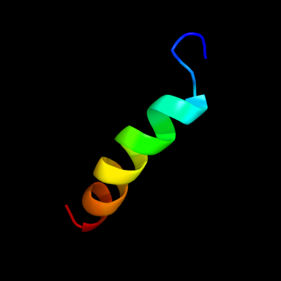
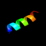
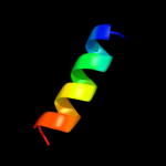
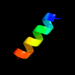
| 1 |
|
PDB 2k21 chain A
Region: 2 - 23
Aligned: 22
Modelled: 22
Confidence: 11.7%
Identity: 18%
PDB header:membrane protein
Chain: A: PDB Molecule:potassium voltage-gated channel subfamily e
PDBTitle: nmr structure of human kcne1 in lmpg micelles at ph 6.0 and2 40 degree c
Phyre2
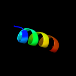
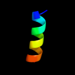
| 2 |
|
PDB 1pd7 chain B
Region: 1 - 9
Aligned: 9
Modelled: 9
Confidence: 11.1%
Identity: 56%
PDB header:transcription
Chain: B: PDB Molecule:mad1;
PDBTitle: extended sid of mad1 bound to the pah2 domain of msin3b
Phyre2
| 3 |
|
PDB 3a0h chain T
Region: 5 - 19
Aligned: 15
Modelled: 15
Confidence: 8.2%
Identity: 47%
PDB header:electron transport
Chain: T: PDB Molecule:photosystem ii reaction center protein t;
PDBTitle: crystal structure of i-substituted photosystem ii complex
Phyre2
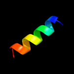
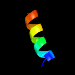
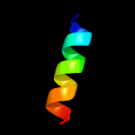
| 4 |
|
PDB 2axt chain T
Region: 5 - 19
Aligned: 15
Modelled: 15
Confidence: 8.2%
Identity: 47%
PDB header:electron transport
Chain: T: PDB Molecule:photosystem ii reaction center t protein;
PDBTitle: crystal structure of photosystem ii from thermosynechococcus elongatus
Phyre2
| 5 |
|
PDB 2axt chain T domain 1
Region: 5 - 19
Aligned: 15
Modelled: 15
Confidence: 8.2%
Identity: 47%
Fold: Single transmembrane helix
Superfamily: Photosystem II reaction center protein T, PsbT
Family: PsbT-like
Phyre2
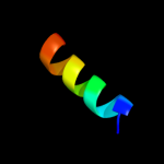
| 6 |
|
PDB 2axt chain T
Region: 5 - 19
Aligned: 15
Modelled: 15
Confidence: 8.2%
Identity: 47%
PDB header:electron transport
Chain: T: PDB Molecule:photosystem ii reaction center t protein;
PDBTitle: crystal structure of photosystem ii from thermosynechococcus elongatus
Phyre2
| 7 |
|
PDB 3kzi chain T
Region: 5 - 19
Aligned: 15
Modelled: 15
Confidence: 8.2%
Identity: 47%
PDB header:electron transport
Chain: T: PDB Molecule:photosystem ii reaction center protein t;
PDBTitle: crystal structure of monomeric form of cyanobacterial photosystem ii
Phyre2
| 8 |
|
PDB 3a0h chain T
Region: 5 - 19
Aligned: 15
Modelled: 15
Confidence: 8.2%
Identity: 47%
PDB header:electron transport
Chain: T: PDB Molecule:photosystem ii reaction center protein t;
PDBTitle: crystal structure of i-substituted photosystem ii complex
Phyre2
| 9 |
|
PDB 3arc chain T
Region: 5 - 19
Aligned: 15
Modelled: 15
Confidence: 8.2%
Identity: 47%
PDB header:electron transport, photosynthesis
Chain: T: PDB Molecule:photosystem ii reaction center protein t;
PDBTitle: crystal structure of oxygen-evolving photosystem ii at 1.9 angstrom2 resolution
Phyre2
| 10 |
|
PDB 3a0b chain T
Region: 5 - 19
Aligned: 15
Modelled: 15
Confidence: 8.2%
Identity: 47%
PDB header:electron transport
Chain: T: PDB Molecule:photosystem ii reaction center protein t;
PDBTitle: crystal structure of br-substituted photosystem ii complex
Phyre2
| 11 |
|
PDB 1s5l chain T
Region: 5 - 19
Aligned: 15
Modelled: 15
Confidence: 8.2%
Identity: 47%
PDB header:photosynthesis
Chain: T: PDB Molecule:photosystem ii psbt protein;
PDBTitle: architecture of the photosynthetic oxygen evolving center
Phyre2
| 12 |
|
PDB 1s5l chain T
Region: 5 - 19
Aligned: 15
Modelled: 15
Confidence: 8.2%
Identity: 47%
PDB header:photosynthesis
Chain: T: PDB Molecule:photosystem ii psbt protein;
PDBTitle: architecture of the photosynthetic oxygen evolving center
Phyre2
| 13 |
|
PDB 3prr chain T
Region: 5 - 19
Aligned: 15
Modelled: 15
Confidence: 8.2%
Identity: 47%
PDB header:photosynthesis
Chain: T: PDB Molecule:photosystem ii reaction center protein t;
PDBTitle: crystal structure of cyanobacterial photosystem ii in complex with2 terbutryn (part 2 of 2). this file contains second monomer of psii3 dimer
Phyre2
| 14 |
|
PDB 3prq chain T
Region: 5 - 19
Aligned: 15
Modelled: 15
Confidence: 8.2%
Identity: 47%
PDB header:photosynthesis
Chain: T: PDB Molecule:photosystem ii reaction center protein t;
PDBTitle: crystal structure of cyanobacterial photosystem ii in complex with2 terbutryn (part 1 of 2). this file contains first monomer of psii3 dimer
Phyre2
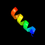
| 15 |
|
PDB 3bz1 chain T
Region: 5 - 19
Aligned: 15
Modelled: 15
Confidence: 8.2%
Identity: 47%
PDB header:electron transport
Chain: T: PDB Molecule:photosystem ii reaction center protein t;
PDBTitle: crystal structure of cyanobacterial photosystem ii (part 12 of 2). this file contains first monomer of psii dimer
Phyre2
| 16 |
|
PDB 3bz2 chain T
Region: 5 - 19
Aligned: 15
Modelled: 15
Confidence: 8.2%
Identity: 47%
PDB header:electron transport
Chain: T: PDB Molecule:photosystem ii reaction center protein t;
PDBTitle: crystal structure of cyanobacterial photosystem ii (part 22 of 2). this file contains second monomer of psii dimer
Phyre2
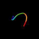
| 17 |
|
PDB 1qgr chain B
Region: 2 - 6
Aligned: 5
Modelled: 5
Confidence: 6.0%
Identity: 60%
PDB header:transport receptor
Chain: B: PDB Molecule:protein (importin alpha-2 subunit);
PDBTitle: structure of importin beta bound to the ibb domain of2 importin alpha (ii crystal form, grown at low ph)
Phyre2