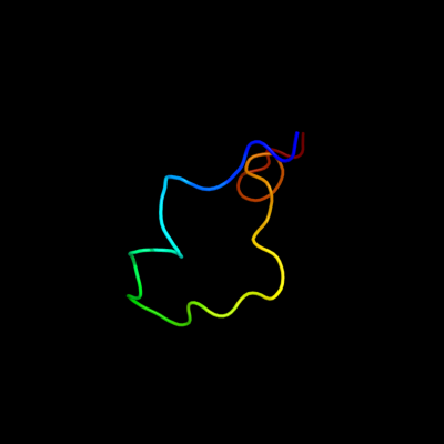
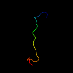
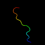
1 c2jtyA_
94.6
39
PDB header: structural proteinChain: A: PDB Molecule: type-1 fimbrial protein, a chain;PDBTitle: self-complemented variant of fima, the main subunit of type 1 pilus
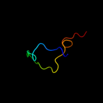
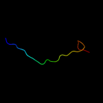
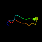
2 c3jwnE_
90.4
20
PDB header: protein binding/cell adhesionChain: E: PDB Molecule: protein fimf;PDBTitle: complex of fimc, fimf, fimg and fimh
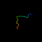
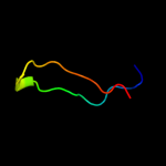
3 c3jwnK_
90.4
20
PDB header: protein binding/cell adhesionChain: K: PDB Molecule: protein fimf;PDBTitle: complex of fimc, fimf, fimg and fimh
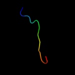
4 c2jmrA_
89.8
20
PDB header: cell adhesionChain: A: PDB Molecule: fimf;PDBTitle: nmr structure of the e. coli type 1 pilus subunit fimf
5 c3jwnL_
89.0
20
PDB header: protein binding/cell adhesionChain: L: PDB Molecule: protein fimf;PDBTitle: complex of fimc, fimf, fimg and fimh
6 c3jwnF_
88.7
20
PDB header: protein binding/cell adhesionChain: F: PDB Molecule: protein fimf;PDBTitle: complex of fimc, fimf, fimg and fimh
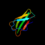
7 d2j2zb1
85.4
26
Fold: Common fold of diphtheria toxin/transcription factors/cytochrome fSuperfamily: Bacterial adhesinsFamily: Pilus subunits8 d2uy6b1
83.4
22
Fold: Common fold of diphtheria toxin/transcription factors/cytochrome fSuperfamily: Bacterial adhesinsFamily: Pilus subunits9 d1cvra1
41.2
18
Fold: Immunoglobulin-like beta-sandwichSuperfamily: E set domainsFamily: Gingipain R (RgpB), C-terminal domain10 c2w07B_
31.7
13
PDB header: cell adhesionChain: B: PDB Molecule: minor pilin subunit papf;PDBTitle: structural determinants of polymerization reactivity of the2 p pilus adaptor subunit papf
11 c1hleB_
31.5
29
PDB header: hydrolase inhibitor(serine proteinase)Chain: B: PDB Molecule: horse leukocyte elastase inhibitor;PDBTitle: crystal structure of cleaved equine leucocyte elastase2 inhibitor determined at 1.95 angstroms resolution
12 d1pdkb_
30.9
24
Fold: Common fold of diphtheria toxin/transcription factors/cytochrome fSuperfamily: Bacterial adhesinsFamily: Pilus subunits13 c1jjoE_
28.7
36
PDB header: signaling proteinChain: E: PDB Molecule: neuroserpin;PDBTitle: crystal structure of mouse neuroserpin (cleaved form)
14 c2h4qB_
28.0
29
PDB header: hydrolase inhibitorChain: B: PDB Molecule: heterochromatin-associated protein ment;PDBTitle: crystal structure of a m-loop deletion variant of ment in2 the cleaved conformation
15 c9paiB_
26.5
46
PDB header: hydrolase inhibitorChain: B: PDB Molecule: protein (plasminogen activator inhibitor-1) residues 365-PDBTitle: cleaved substrate variant of plasminogen activator inhibitor-1
16 d1i8na_
25.7
32
Fold: Hairpin loop containing domain-likeSuperfamily: Hairpin loop containing domain-likeFamily: Anti-platelet protein17 c1i8nA_
25.7
32
PDB header: toxinChain: A: PDB Molecule: anti-platelet protein;PDBTitle: crystal structure of leech anti-platelet protein
18 c2rivB_
22.1
38
PDB header: signaling proteinChain: B: PDB Molecule: thyroxine-binding globulin;PDBTitle: crystal structure of the reactive loop cleaved human thyroxine binding2 globulin
19 c7apiB_
20.7
31
PDB header: proteinase inhibitorChain: B: PDB Molecule: alpha 1-antitrypsin;PDBTitle: the s variant of human alpha1-antitrypsin, structure and implications2 for function and metabolism
20 c3f02C_
20.7
36
PDB header: hydrolase inhibitorChain: C: PDB Molecule: neuroserpin;PDBTitle: cleaved human neuroserpin
21 c1mtpB_
not modelled
16.2
46
PDB header: structural genomicsChain: B: PDB Molecule: serine proteinase inhibitor (serpin), chain b;PDBTitle: the x-ray crystal structure of a serpin from a thermophilic2 prokaryote
22 c3cooB_
not modelled
14.1
26
PDB header: cell adhesionChain: B: PDB Molecule: spondin-1;PDBTitle: the crystal structure of reelin-n domain of f-spondin
23 c3i4oA_
not modelled
13.3
19
PDB header: translationChain: A: PDB Molecule: translation initiation factor if-1;PDBTitle: crystal structure of translation initiation factor 1 from2 mycobacterium tuberculosis
24 c2zouB_
not modelled
11.9
26
PDB header: cell adhesionChain: B: PDB Molecule: spondin-1;PDBTitle: crystal struture of human f-spondin reeler domain (fragment 2)
25 d1exta2
not modelled
8.2
36
Fold: TNF receptor-likeSuperfamily: TNF receptor-likeFamily: TNF receptor-like26 c2j98A_
not modelled
7.5
22
PDB header: rna-binding proteinChain: A: PDB Molecule: replicase polyprotein 1ab;PDBTitle: human coronavirus 229e non structural protein 9 cys69ala2 mutant (nsp9)
27 d1qz8a_
not modelled
7.5
40
Fold: Replicase NSP9Superfamily: Replicase NSP9Family: Replicase NSP928 c1uw7A_
not modelled
7.4
40
PDB header: replicase proteinChain: A: PDB Molecule: nsp9;PDBTitle: nsp9 protein from sars-coronavirus.
29 d1gtka2
not modelled
6.9
13
Fold: dsRBD-likeSuperfamily: Porphobilinogen deaminase (hydroxymethylbilane synthase), C-terminal domainFamily: Porphobilinogen deaminase (hydroxymethylbilane synthase), C-terminal domain30 c1ow1A_
not modelled
6.7
37
PDB header: transcriptionChain: A: PDB Molecule: smart/hdac1 associated repressor protein;PDBTitle: crystal structure of the spoc domain of the human2 transcriptional corepressor, sharp.
31 d1ow1a_
not modelled
6.7
37
Fold: SPOC domain-likeSuperfamily: SPOC domain-likeFamily: SPOC domain32 c2d3jA_
not modelled
6.7
9
PDB header: signaling protein inhibitorChain: A: PDB Molecule: wnt inhibitory factor-1;PDBTitle: nmr structure of the wif domain from human wif-1
33 d1n26a1
not modelled
6.6
22
Fold: Immunoglobulin-like beta-sandwichSuperfamily: ImmunoglobulinFamily: I set domains34 d2ij0c1
not modelled
6.2
24
Fold: Immunoglobulin-like beta-sandwichSuperfamily: ImmunoglobulinFamily: V set domains (antibody variable domain-like)35 c2kc2A_
not modelled
5.8
27
PDB header: structural proteinChain: A: PDB Molecule: talin-1;PDBTitle: nmr structure of the f1 domain (residues 86-202) of the2 talin
36 c3m6wA_
not modelled
5.7
20
PDB header: transferaseChain: A: PDB Molecule: rrna methylase;PDBTitle: multi-site-specific 16s rrna methyltransferase rsmf from thermus2 thermophilus in space group p21212 in complex with s-adenosyl-l-3 methionine
37 c2frxD_
not modelled
5.6
20
PDB header: transferaseChain: D: PDB Molecule: hypothetical protein yebu;PDBTitle: crystal structure of yebu, a m5c rna methyltransferase from e.coli
38 d1ikpa1
not modelled
5.5
17
Fold: Concanavalin A-like lectins/glucanasesSuperfamily: Concanavalin A-like lectins/glucanasesFamily: Exotoxin A, N-terminal domain39 c2dllA_
not modelled
5.3
10
PDB header: cytokineChain: A: PDB Molecule: interferon regulatory factor 4;PDBTitle: solution structure of the irf domain of human interferon2 regulator factors 4
40 d1pdaa2
not modelled
5.2
17
Fold: dsRBD-likeSuperfamily: Porphobilinogen deaminase (hydroxymethylbilane synthase), C-terminal domainFamily: Porphobilinogen deaminase (hydroxymethylbilane synthase), C-terminal domain41 d3c2ah1
not modelled
5.1
20
Fold: Immunoglobulin-like beta-sandwichSuperfamily: ImmunoglobulinFamily: V set domains (antibody variable domain-like)