| 1 |
|
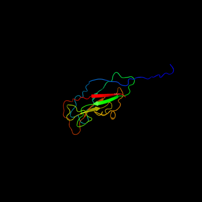
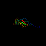
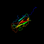
PDB 3jwn chain L
Region: 31 - 170
Aligned: 139
Modelled: 140
Confidence: 99.9%
Identity: 19%
PDB header:protein binding/cell adhesion
Chain: L: PDB Molecule:protein fimf;
PDBTitle: complex of fimc, fimf, fimg and fimh
Phyre2
| 2 |
|
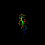
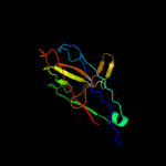
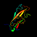
PDB 3jwn chain K
Region: 31 - 170
Aligned: 139
Modelled: 140
Confidence: 99.9%
Identity: 19%
PDB header:protein binding/cell adhesion
Chain: K: PDB Molecule:protein fimf;
PDBTitle: complex of fimc, fimf, fimg and fimh
Phyre2
| 3 |
|
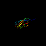
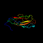
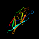
PDB 3jwn chain E
Region: 31 - 170
Aligned: 139
Modelled: 140
Confidence: 99.9%
Identity: 19%
PDB header:protein binding/cell adhesion
Chain: E: PDB Molecule:protein fimf;
PDBTitle: complex of fimc, fimf, fimg and fimh
Phyre2
| 4 |
|
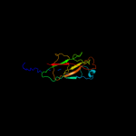
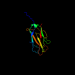
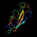
PDB 3jwn chain F
Region: 31 - 170
Aligned: 139
Modelled: 140
Confidence: 99.9%
Identity: 19%
PDB header:protein binding/cell adhesion
Chain: F: PDB Molecule:protein fimf;
PDBTitle: complex of fimc, fimf, fimg and fimh
Phyre2
| 5 |
|
PDB 2w07 chain B
Region: 41 - 169
Aligned: 117
Modelled: 129
Confidence: 99.9%
Identity: 27%
PDB header:cell adhesion
Chain: B: PDB Molecule:minor pilin subunit papf;
PDBTitle: structural determinants of polymerization reactivity of the2 p pilus adaptor subunit papf
Phyre2
| 6 |
|
PDB 2jmr chain A
Region: 31 - 170
Aligned: 139
Modelled: 140
Confidence: 99.9%
Identity: 18%
PDB header:cell adhesion
Chain: A: PDB Molecule:fimf;
PDBTitle: nmr structure of the e. coli type 1 pilus subunit fimf
Phyre2
| 7 |
|
PDB 2uy6 chain B domain 1
Region: 35 - 170
Aligned: 136
Modelled: 136
Confidence: 99.9%
Identity: 20%
Fold: Common fold of diphtheria toxin/transcription factors/cytochrome f
Superfamily: Bacterial adhesins
Family: Pilus subunits
Phyre2
| 8 |
|
PDB 2j2z chain B domain 1
Region: 35 - 170
Aligned: 135
Modelled: 136
Confidence: 99.9%
Identity: 18%
Fold: Common fold of diphtheria toxin/transcription factors/cytochrome f
Superfamily: Bacterial adhesins
Family: Pilus subunits
Phyre2
| 9 |
|
PDB 2jty chain A
Region: 26 - 170
Aligned: 143
Modelled: 145
Confidence: 99.9%
Identity: 20%
PDB header:structural protein
Chain: A: PDB Molecule:type-1 fimbrial protein, a chain;
PDBTitle: self-complemented variant of fima, the main subunit of type 1 pilus
Phyre2
| 10 |
|
PDB 3bfw chain A
Region: 44 - 169
Aligned: 124
Modelled: 126
Confidence: 99.8%
Identity: 11%
PDB header:structural protein/structural protein
Chain: A: PDB Molecule:protein fimg;
PDBTitle: crystal structure of truncated fimg (fimgt) in complex with the donor2 strand peptide of fimf (dsf)
Phyre2
| 11 |
|
PDB 1n12 chain A
Region: 45 - 169
Aligned: 125
Modelled: 125
Confidence: 99.8%
Identity: 18%
Fold: Common fold of diphtheria toxin/transcription factors/cytochrome f
Superfamily: Bacterial adhesins
Family: Pilus subunits
Phyre2
| 12 |
|
PDB 1pdk chain B
Region: 41 - 170
Aligned: 130
Modelled: 130
Confidence: 99.8%
Identity: 15%
Fold: Common fold of diphtheria toxin/transcription factors/cytochrome f
Superfamily: Bacterial adhesins
Family: Pilus subunits
Phyre2
| 13 |
|
PDB 3bwu chain F
Region: 58 - 170
Aligned: 112
Modelled: 113
Confidence: 99.7%
Identity: 20%
PDB header:chaperone, structural, membrane protein
Chain: F: PDB Molecule:protein fimf;
PDBTitle: crystal structure of the ternary complex of fimd (n-terminal domain,2 fimdn) with fimc and the n-terminally truncated pilus subunit fimf3 (fimft)
Phyre2
| 14 |
|
PDB 1ze3 chain H domain 1
Region: 46 - 170
Aligned: 114
Modelled: 125
Confidence: 99.7%
Identity: 18%
Fold: Common fold of diphtheria toxin/transcription factors/cytochrome f
Superfamily: Bacterial adhesins
Family: Pilus subunits
Phyre2
| 15 |
|
PDB 1klf chain P
Region: 34 - 170
Aligned: 126
Modelled: 129
Confidence: 99.7%
Identity: 17%
PDB header:chaperone/adhesin complex
Chain: P: PDB Molecule:fimh protein;
PDBTitle: fimh adhesin-fimc chaperone complex with d-mannose
Phyre2
| 16 |
|
PDB 3qbt chain H
Region: 45 - 111
Aligned: 61
Modelled: 67
Confidence: 28.9%
Identity: 15%
PDB header:protein transport/hydrolase
Chain: H: PDB Molecule:inositol polyphosphate 5-phosphatase ocrl-1;
PDBTitle: crystal structure of ocrl1 540-678 in complex with rab8a:gppnhp
Phyre2