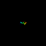
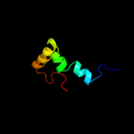
PDB header:ribosome Chain: 2: PDB Molecule:50s ribosomal protein l34; PDBTitle: structures of the bacterial ribosome in classical and hybrid states of2 trna binding
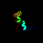
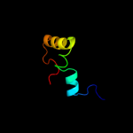
Confidence and coverage
Confidence:
99.9%
Coverage:
100%
46 residues (100% of your sequence) have been modelled with 99.9% confidence by the single highest scoring template.
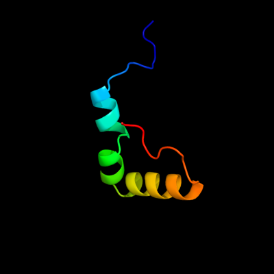
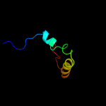
Region: 1 - 46 Aligned: 46 Modelled: 46 Confidence: 99.9% Identity: 100% PDB header:ribosome Chain: E: PDB Molecule:50s ribosomal protein l3; PDBTitle: structural insights into cognate vs. near-cognate discrimination2 during decoding. this entry contains the large subunit of a ribosome3 programmed with a cognate codon
Region: 1 - 46 Aligned: 46 Modelled: 46 Confidence: 99.9% Identity: 100% PDB header:ribosome Chain: E: PDB Molecule:50s ribosomal protein l3; PDBTitle: structural insights into cognate vs. near-cognate discrimination2 during decoding. this entry contains the large subunit of a ribosome3 programmed with a near-cognate codon.
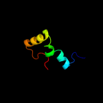
Region: 1 - 46 Aligned: 46 Modelled: 46 Confidence: 99.9% Identity: 100% PDB header:ribosome Chain: 2: PDB Molecule:50s ribosomal protein l34; PDBTitle: crystal structure of the bacterial ribosome from2 escherichia coli in complex with gentamicin. this file3 contains the 50s subunit of the second 70s ribosome, with4 gentamicin bound. the entire crystal structure contains5 two 70s ribosomes and is described in remark 400.
Region: 1 - 46 Aligned: 46 Modelled: 46 Confidence: 99.9% Identity: 100% PDB header:ribosome Chain: 2: PDB Molecule:50s ribosomal protein l34; PDBTitle: crystal structure of the bacterial ribosome from2 escherichia coli in complex with neomycin. this file3 contains the 50s subunit of the second 70s ribosome, with4 neomycin bound. the entire crystal structure contains two5 70s ribosomes and is described in remark 400.
Region: 1 - 46 Aligned: 46 Modelled: 46 Confidence: 99.9% Identity: 100% PDB header:ribosome Chain: 2: PDB Molecule:50s ribosomal protein l34; PDBTitle: crystal structure of the bacterial ribosome from2 escherichia coli in complex with gentamicin. this file3 contains the 50s subunit of the first 70s ribosome, with4 gentamicin bound. the entire crystal structure contains5 two 70s ribosomes and is described in remark 400.
Region: 1 - 46 Aligned: 46 Modelled: 46 Confidence: 99.9% Identity: 100% PDB header:ribosome Chain: 2: PDB Molecule:50s ribosomal protein l34; PDBTitle: crystal structure of the bacterial ribosome from2 escherichia coli in complex with neomycin. this file3 contains the 50s subunit of the first 70s ribosome, with4 neomycin bound. the entire crystal structure contains two5 70s ribosomes and is described in remark 400.
Region: 1 - 46 Aligned: 46 Modelled: 46 Confidence: 99.9% Identity: 100% PDB header:ribosome Chain: 2: PDB Molecule:50s ribosomal protein l34; PDBTitle: crystal structure of the bacterial ribosome from escherichia2 coli in complex with paromomycin and ribosome recycling3 factor (rrf). this file contains the 50s subunit of the4 second 70s ribosome, with paromomycin and rrf bound. the5 entire crystal structure contains two 70s ribosomes and is6 described in remark 400.
Region: 1 - 46 Aligned: 46 Modelled: 46 Confidence: 99.9% Identity: 100% PDB header:ribosome Chain: 2: PDB Molecule:50s ribosomal protein l34; PDBTitle: crystal structure of the bacterial ribosome from escherichia2 coli in complex with paromomycin and ribosome recycling3 factor (rrf). this file contains the 50s subunit of the4 first 70s ribosome, with paromomycin and rrf bound. the5 entire crystal structure contains two 70s ribosomes and is6 described in remark 400.
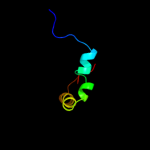
Region: 1 - 46 Aligned: 46 Modelled: 46 Confidence: 99.9% Identity: 100% PDB header:ribosome Chain: V: PDB Molecule:50s ribosomal protein l34; PDBTitle: structure of the 50s subunit of e. coli ribosome in post-2 accommodation state
Region: 1 - 46 Aligned: 46 Modelled: 46 Confidence: 99.9% Identity: 100% PDB header:ribosome Chain: 2: PDB Molecule:50s ribosomal protein l34; PDBTitle: crystal structure of the bacterial ribosome from escherichia2 coli in complex with ribosome recycling factor (rrf). this3 file contains the 50s subunit of the first 70s ribosome,4 with rrf bound. the entire crystal structure contains two5 70s ribosomes and is described in remark 400.
Region: 1 - 46 Aligned: 46 Modelled: 46 Confidence: 99.9% Identity: 100% PDB header:ribosome Chain: 2: PDB Molecule:50s ribosomal protein l34; PDBTitle: crystal structure of the bacterial ribosome from escherichia2 coli in complex with ribosome recycling factor (rrf). this3 file contains the 50s subunit of the second 70s ribosome,4 with rrf bound. the entire crystal structure contains two5 70s ribosomes and is described in remark 400.
Region: 1 - 46 Aligned: 46 Modelled: 46 Confidence: 99.9% Identity: 100% PDB header:ribosome Chain: 2: PDB Molecule:50s ribosomal protein l34; PDBTitle: crystal structure of the bacterial ribosome from escherichia2 coli in complex with spectinomycin and neomycin. this file3 contains the 50s subunit of the second 70s ribosome, with4 neomycin bound. the entire crystal structure contains two5 70s ribosomes.
Region: 1 - 46 Aligned: 46 Modelled: 46 Confidence: 99.9% Identity: 100% PDB header:ribosome Chain: 2: PDB Molecule:50s ribosomal protein l34; PDBTitle: crystal structure of the bacterial ribosome from escherichia2 coli in complex with spectinomycin and neomycin. this file3 contains the 50s subunit of the first 70s ribosome, with4 neomycin bound. the entire crystal structure contains two5 70s ribosomes.
Region: 1 - 46 Aligned: 46 Modelled: 46 Confidence: 99.9% Identity: 100% PDB header:ribosome Chain: 2: PDB Molecule:50s ribosomal protein l34; PDBTitle: crystal structure of the bacterial ribosome from escherichia2 coli in complex with gentamicin and ribosome recycling3 factor (rrf). this file contains the 50s subunit of the4 first 70s ribosome, with gentamicin and rrf bound. the5 entire crystal structure contains two 70s ribosomes and is6 described in remark 400.
Region: 1 - 46 Aligned: 46 Modelled: 46 Confidence: 99.9% Identity: 100% PDB header:ribosome Chain: 2: PDB Molecule:50s ribosomal protein l34; PDBTitle: crystal structure of the bacterial ribosome from escherichia2 coli in complex with gentamicin and ribosome recycling3 factor (rrf). this file contains the 50s subunit of the4 second 70s ribosome, with gentamicin and rrf bound. the5 entire crystal structure contains two 70s ribosomes and is6 described in remark 400.
Phyre2
21
22
23
24
25
26
27
28
29
30
31
32
33
34
35
36
37
38
39
40
41
42
43
44
45
46
47
48
49
50
51
52
53
54
55
56
Detailed template information
Binding site prediction
Due to computational demand, binding site predictions are not run for batch jobs
If you want to predict binding sites, please manually submit your model of choice to 3DLigandSite
Phyre is for academic use only
Please cite: Protein structure prediction on
the web: a case study using the Phyre server
Kelley LA and Sternberg MJE. Nature Protocols
4, 363 - 371 (2009) [pdf] [Import into BibTeX]
If you use the binding site
predictions from 3DLigandSite, please also cite:
3DLigandSite: predicting ligand-binding sites using similar structures.
Wass MN, Kelley LA and Sternberg
MJ Nucleic Acids Research 38, W469-73 (2010) [PubMed]